High tibial osteotomy is a surgical procedure performed to relieve pressure on the damaged site of an arthritic knee joint. It is usually performed in arthritic conditions affecting only one side of your knee and the aim is to take pressure off the damaged area and shift it to the other side of your knee with healthy cartilage. During the surgery, your surgeon will remove or add a wedge of bone either below or above the knee joint depending on the site of arthritic damage.
High tibial osteotomy is commonly used for patients with osteoarthritis that is isolated to a single compartment (unicompartmental osteoarthritis). It is also performed for treating a variety of knee conditions such as gonarthrosis with varus or valgus malalignment, osteochondritis dissecans, osteonecrosis, posterolateral instability, and chondral resurfacing.
The goal of the surgery is to release the involved joint compartment by correcting the malalignment of the tibia and to maintain the joint line perpendicular to the mechanical axis of the leg. There are two techniques that may be used: closing wedge osteotomy and opening wedge osteotomy. The surgeon determines the choice of the technique based on the requirement of the patient.
Closing wedge osteotomy is the most commonly used technique to perform high tibial osteotomy. In this procedure, the surgeon makes an incision in front of the knee and removes a small wedge of bone from the upper part of the tibia or shinbone. This manipulation brings the bones together and fills the space left by the removed bone. The surgeon then uses plates and screws to bind the bones together while the osteotomy heals. This procedure unloads the pressure off the damaged joint area and helps to transfer some of the weight to the outer part of the knee, where the cartilage is still intact.
In this procedure, the surgeon makes an incision in front of the knee, just below the kneecap and makes a wedge-shaped cut in the bone. Bone graft is used to fill the space of the wedge-shaped opening and if required plates and screws can be attached to further support the surgical site during the healing process. This realignment increases the angle of the knee to relieve the painful symptoms.
Complications following high tibial osteotomy may include infection, skin necrosis, non-union (failure of the bones to heal), nerve injury, blood vessel injury, failure to correct the varus deformity, compartment syndrome and deep vein thrombosis or blood clots.
During knee replacement surgery, damaged bone and cartilage are resurfaced with metal and plastic components. In unicompartmental knee replacement (also called “partial” knee replacement) only a portion of the knee is resurfaced. This procedure is an alternative to total knee replacement for patients whose disease is limited to just one area of the knee. Because a partial knee replacement is done through a smaller incision, patients usually spend less time in the hospital and return to normal activities sooner than total knee replacement patients. There is a range of treatments for knee osteoarthritis and your doctor will discuss with you the options that will best relieve your individual osteoarthritis symptoms.
In a unicompartmental knee replacement, only one area of the knee is resurfaced.
In knee osteoarthritis, the cartilage protecting the bones of the knee slowly wears away. This can occur throughout the knee joint or just in a single area of the knee.
(Left) A normal knee joint: The medial, lateral, and patellofemoral compartments are shown with red arrows. (Right) An x-ray of a normal knee joint shows healthy space between the bones. Advanced osteoarthritis that is limited to a single compartment may be treated with a unicompartmental knee replacement. During this procedure, the damaged compartment is replaced with metal and plastic. The healthy cartilage and bone, as well as all of the ligaments, are preserved.
(Left) Osteoarthritis is limited to the medial compartment. (Right) This x-ray shows severe osteoarthritis with “bone-on-bone” degeneration in the medial compartment (arrow).
Multiple studies show that a majority of patients who are appropriate candidates for the procedure have good results with a unicompartmental knee replacement.
The advantages of partial knee replacement over total knee replacement include:
Also, because the bone, cartilage, and ligaments in the healthy parts of the knee are kept, many patients report that a unicompartmental knee replacement feels more natural than a total knee replacement. A unicompartmental knee may also bend better.
The disadvantages of partial knee replacement compared with total knee replacement include:
An advantage of partial knee replacement over total knee replacement is that healthy parts of the knee are preserved, which helps to maintain a more “natural” function of the knee.
If your osteoarthritis has advanced and nonsurgical treatment options are no longer relieving your symptoms, your doctor may recommend knee replacement surgery.To be a candidate for unicompartmental knee replacement, your arthritis must be limited to one compartment of your knee. In addition, if you have any of the following characteristics, you may not be eligible for the procedure:
With the proper patient selection, modern unicompartmental knee replacements have demonstrated excellent medium- and long-term results in both younger and older patients.
A thorough evaluation with an orthopedic surgeon will determine whether you are a good candidate for a partial knee replacement.
Your doctor will ask you several questions about your general health, your knee pain, and your ability to function.
He or she will be specifically concerned with the location of your pain. If your pain is located almost entirely on either the inside portion or outside portion of your knee, then you may be a candidate for a partial knee replacement. If you have pain throughout your entire knee or pain in the front of your knee (under your kneecap) you may be better qualified for a total knee replacement.
Your doctor will closely examine your knee. He or she will try to determine the location of your pain. Your doctor will also test your knee for range of motion and ligament quality. If your knee is too stiff, or if the ligaments in your knee feel weak or torn, then your doctor will probably not recommend unicompartmental knee replacement (although you still may be a great candidate for a total knee replacement).
You will likely be admitted to the hospital on the day of surgery. Before your procedure, a doctor from the anesthesia department will discuss anesthesia choices with you. You should also have discussed anesthesia choices with your surgeon during your preoperative clinic visits. Anesthesia options include:
Your surgeon will also see you before surgery and sign your knee to verify the surgical site.
A partial knee replacement operation typically lasts between 1 and 2 hours. Inspection of the joint. Your surgeon will make an incision at the front of your knee. He or she will then explore the three compartments of your knee to verify that the cartilage damage is, in fact, limited to one compartment and that your ligaments are intact. If your surgeon feels that your knee is unsuitable for a partial knee replacement, he or she may instead perform a total knee replacement. This contingency plan will have been discussed with you before your operation to make sure that you agree with this strategy.
X-rays of a good candidate for partial knee replacement. (Left) Severe osteoarthritis is limited to the medial compartment. (Right) The same knee after partial knee replacement.
There are three basic steps in the procedure:
Recovery room. After the surgery, you will be taken to the recovery room, where you will be closely monitored by nurses as you recover from the anesthesia. You will then be taken to your hospital room.
As with any surgical procedure, there are risks involved with a partial knee replacement. Your surgeon will discuss each of the risks with you and will take specific measures to help avoid potential complications.
Although rare, the most common risks include:
Hospital discharge. Partial knee replacement patients usually experience less postoperative pain, less swelling, and have easier rehabilitation than patients undergoing total knee replacement. In most cases, patients go home 1 to 3 days after the operation. Some patients go home the day of the surgery.
After surgery, you will feel some pain, but your surgeon and nurses will make every effort to help you feel as comfortable as possible. Many types of medicines are available to help control pain, including opioids, non-steroidal anti-inflammatory drugs (NSAIDs), and local
Treating pain with medication can help you feel more comfortable, which will help your body heal and recover from surgery faster.
Opioids can provide excellent pain relief, however, they are a narcotic and can be addictive. It is important to use opioids only as directed by your doctor. You should stop taking these medications as soon as your pain starts to improve.
You will begin putting weight on your knee immediately after surgery. You may need a walker, cane, or crutches for the first several days or weeks until you become comfortable enough to walk without assistance.
A physical therapist will give you exercises to help maintain your range of motion and restore your strength.
You will continue to see your orthopedic surgeon for follow-up visits in his or her clinic at regular intervals. You will most likely resume all of your regular activities of daily living by 6 weeks after surgery.
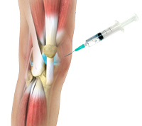
Synvisc is one of the most commonly used hyaluronan preparations. It is indicated in the management of shoulder, knee, hip, or ankle osteoarthritis that has not responded to non-surgical treatment options such as pain medications, physical therapy, and corticosteroid injections.
Synvisc provides symptomatic relief and delays the need for surgery. It is injected directly into the joint to replenish the diminished synovial fluid, thereby enhancing its lubricating properties. A single dose or a total of three separate doses of Synvisc, over several weeks, may be required for optimum benefit.
Synvisc injection not only supplements the hyaluronan in the joint but also stimulates the production of hyaluronan in the treated knee. This provides gradual symptomatic relief over the course of the injections. This effect may last for several months.
Ice packs and an analgesic may be used, if required, to ease the discomfort. Any strenuous activity such as jogging or tennis should be completely avoided for 48 hours to a week after the injection and should be resumed only after consultation with your doctor.
You may experience mild pain, swelling, warmth, and redness at the injection site for up to 48 hours following a Synvisc injection. Headache and joint stiffness may also occur in some cases.
Immediately consult your doctor if you develop fever or the pain and swelling fail to resolve after 48 hours following the injection.