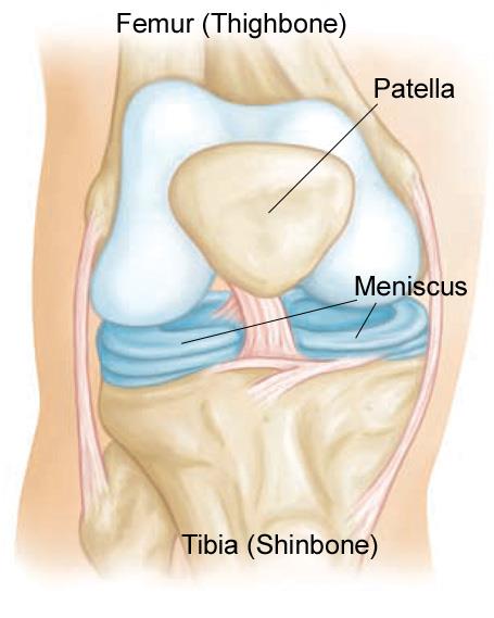
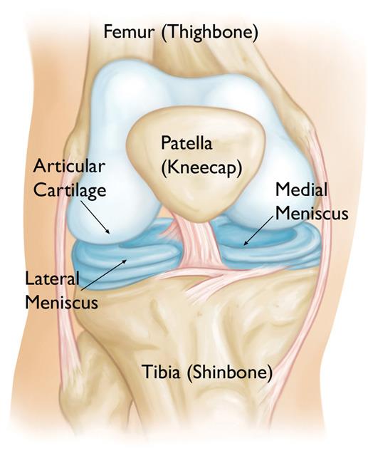
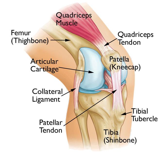
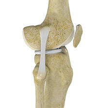
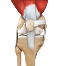
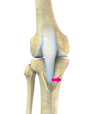
Three bones meet to form your knee joint: your thighbone (femur), shinbone (tibia), and kneecap (patella). Your kneecap sits in front of the joint to provide some protection.
Bones are connected to other bones by ligaments. There are four primary ligaments in your knee. They act like strong ropes to hold the bones together and keep your knee stable.
These are found on the sides of your knee. The medial collateral ligament is on the inside and the lateral collateral ligament is on the outside. They control the sideways motion of your knee and brace it against unusual movement.
These are found inside your knee joint. They cross each other to form an “X” with the anterior cruciate ligament in front and the posterior cruciate ligament in back. The cruciate ligaments control the back and forth motion of your knee.
The anterior cruciate ligament runs diagonally in the middle of the knee. It prevents the tibia from sliding out in front of the femur, as well as provides rotational stability to the knee.
About half of all injuries to the anterior cruciate ligament occur along with damage to other structures in the knee, such as articular cartilage, meniscus, or other ligaments.
Injured ligaments are considered “sprains” and are graded on a severity scale.
Grade 1 Sprains. The ligament is mildly damaged in a Grade 1 Sprain. It has been slightly stretched, but is still able to help keep the knee joint stable.
Grade 2 Sprains. A Grade 2 Sprain stretches the ligament to the point where it becomes loose. This is often referred to as a partial tear of the ligament.
Grade 3 Sprains. This type of sprain is most commonly referred to as a complete tear of the ligament. The ligament has been split into two pieces, and the knee joint is unstable.
Partial tears of the anterior cruciate ligament are rare; most ACL injuries are complete or near complete tears.
The anterior cruciate ligament can be injured in several ways:
Several studies have shown that female athletes have a higher incidence of ACL injury than male athletes in certain sports. It has been proposed that this is due to differences in physical conditioning, muscular strength, and neuromuscular control. Other suggested causes include differences in pelvis and lower extremity (leg) alignment, increased looseness in ligaments, and the effects of estrogen on ligament properties.
When you injure your anterior cruciate ligament, you might hear a “popping” noise and you may feel your knee give out from under you. Other typical symptoms include:
During your first visit, your doctor will talk to you about your symptoms and medical history.
During the physical examination, your doctor will check all the structures of your injured knee, and compare them to your non-injured knee. Most ligament injuries can be diagnosed with a thorough physical examination of the knee.
Other tests which may help your doctor confirm your diagnosis include:
X-rays. Although they will not show any injury to your anterior cruciate ligament, x-rays can show whether the injury is associated with a broken bone.
Magnetic resonance imaging (MRI) scan. This study creates better images of soft tissues like the anterior cruciate ligament. However, an MRI is usually not required to make the diagnosis of a torn ACL.
Treatment for an ACL tear will vary depending upon the patient’s individual needs. For example, the young athlete involved in agility sports will most likely require surgery to safely return to sports. The less active, usually older, individual may be able to return to a quieter lifestyle without surgery.
A torn ACL will not heal without surgery. But nonsurgical treatment may be effective for patients who are elderly or have a very low activity level. If the overall stability of the knee is intact, your doctor may recommend simple, nonsurgical options.
Bracing. Your doctor may recommend a brace to protect your knee from instability. To further protect your knee, you may be given crutches to keep you from putting weight on your leg.
Rebuilding the ligament. Most ACL tears cannot be sutured (stitched) back together. To surgically repair the ACL and restore knee stability, the ligament must be reconstructed. Your doctor will replace your torn ligament with a tissue graft. This graft acts as a scaffolding for a new ligament to grow on.
Grafts can be obtained from several sources. Often they are taken from the patellar tendon, which runs between the kneecap and the shinbone. Hamstring tendons at the back of the thigh are a common source of grafts. Sometimes a quadriceps tendon, which runs from the kneecap into the thigh, is used. Finally, cadaver graft (allograft) can be used.
There are advantages and disadvantages to all graft sources. You should discuss graft choices with your own orthopaedic surgeon to help determine which is best for you.
Because the regrowth takes time, it may be six months or more before an athlete can return to sports after surgery.
Procedure. Surgery to rebuild an anterior cruciate ligament is done with an arthroscope using small incisions. Arthroscopic surgery is less invasive. The benefits of less invasive techniques include less pain from surgery, less time spent in the hospital, and quicker recovery times.
Whether your treatment involves surgery or not, rehabilitation plays a vital role in getting you back to your daily activities. A physical therapy program will help you regain knee strength and motion.
If you have surgery, physical therapy first focuses on returning motion to the joint and surrounding muscles. This is followed by a strengthening program designed to protect the new ligament. This strengthening gradually increases the stress across the ligament. The final phase of rehabilitation is aimed at a functional return tailored for the athlete’s sport.
Two bones meet to form your knee joint: your thighbone (femur) and shinbone (tibia). Your kneecap sits in front of the joint to provide some protection.
Bones are connected to other bones by ligaments. There are four primary ligaments in your knee. They act like strong ropes to hold the bones together and keep your knee stable.
Collateral ligaments. These are found on the sides of your knee. The medial collateral ligament is on the inside and the lateral collateral ligament is on the outside. They control the sideways motion of your knee and brace it against unusual movement.
Cruciate ligaments. These are found inside your knee joint. They cross each other to form an “X” with the anterior cruciate ligament in front and the posterior cruciate ligament in back. The cruciate ligaments control the back and forth motion of your knee.
The posterior cruciate ligament keeps the shinbone from moving backwards too far. It is stronger than the anterior cruciate ligament and is injured less often. The posterior cruciate ligament has two parts that blend into one structure that is about the size of a person’s little finger.
Injuries to the posterior cruciate ligament are not as common as other knee ligament injuries. In fact, they are often subtle and more difficult to evaluate than other ligament injuries in the knee.
Many times a posterior cruciate ligament injury occurs along with injuries to other structures in the knee such as cartilage, other ligaments, and bone.
Injured ligaments are considered “sprains” and are graded on a severity scale.
Grade 1 Sprains. The ligament is mildly damaged in a Grade 1 Sprain. It has been slightly stretched, but is still able to help keep the knee joint stable.
Grade 2 Sprains. A Grade 2 Sprain stretches the ligament to the point where it becomes loose. This is often referred to as a partial tear of the ligament.
Grade 3 Sprains. This type of sprain is most commonly referred to as a complete tear of the ligament. The ligament has been split into two pieces, and the knee joint is unstable.
Posterior cruciate ligament tears tend to be partial tears with the potential to heal on their own. People who have injured just their posterior cruciate ligaments are usually able to return to sports without knee stability problems.
An injury to the posterior cruciate ligament can happen many ways. It typically requires a powerful force.
The typical symptoms of a posterior cruciate ligament injury are:
During your first visit, your doctor will talk to you about your symptoms and medical history.
During the physical examination, your doctor will check all the structures of your injured knee, and compare them to your non-injured knee. Your injured knee may appear to sag backwards when bent. It might slide backwards too far, particularly when it is bent beyond a 90° angle. Other tests which may help your doctor confirm your diagnosis include X-rays and magnetic resonance imaging (MRI). It is possible, however, for these pictures to appear normal, especially if the injury occurred more than 3 months before the tests.
X-rays. Although they will not show any injury to your posterior cruciate ligament, X-rays can show whether the ligament tore off a piece of bone when it was injured. This is called an avulsion fracture.
MRI. This study creates better images of soft tissues like the posterior cruciate ligament
If you have injured just your posterior cruciate ligament, your injury may heal quite well without surgery Your doctor may recommend simple, nonsurgical options.
RICE. When you are first injured, the RICE method – rest, ice, gentle compression and elevation — can help speed your recovery.
Immobilization. Your doctor may recommend a brace to prevent your knee from moving. To further protect your knee, you may be given crutches to keep you from putting weight on your leg.
Physical therapy. As the swelling goes down, a careful rehabilitation program is started. Specific exercises will restore function to your knee and strengthen the leg muscles that support it. Strengthening the muscles in the front of your thigh (quadriceps) has been shown to be a key factor in a successful recovery.
Your doctor may recommend surgery if you have combined injuries. For example, if you have dislocated your knee and torn multiple ligaments including the posterior cruciate ligament, surgery is almost always necessary.
Rebuilding the ligament. Because sewing the ligament ends back together does not usually heal, a torn posterior cruciate ligament must be rebuilt. Your doctor will replace your torn ligament with a tissue graft. This graft is taken from another part of your body, or from another human donor (cadaver). It can take several months for the graft to heal into your bone.
Procedure. Surgery to rebuild a posterior cruciate ligament is done with an arthroscope using small incisions. Arthroscopic surgery is less invasive. The benefits of less invasive techniques include less pain from surgery, less time spent in the hospital, and quicker recovery times.
Surgical procedures to repair posterior cruciate ligaments continue to improve. More advanced techniques help patients resume a wider range of activities after rehabilitation.
Whether your treatment involves surgery or not, rehabilitation plays a vital role in getting you back to your daily activities. A physical therapy program will help you regain knee strength and motion. If you had surgery, physical therapy will begin 1 to 4 weeks after your procedure.
How long it takes you to recover from a posterior cruciate ligament injury will depend on the severity of your injury. Combined injuries often have a slow recovery, but most patients do well over time.
If your injury requires surgery, it may be several weeks before you return to a desk job – perhaps months if your job requires a lot of activity. Full recovery typically requires 6 to 12 months.
Although it is a slow process, your commitment to therapy is the most important factor in returning to all the activities you enjoy.
.
Three bones meet to form your knee joint: your thighbone (femur), shinbone (tibia), and kneecap (patella).
Two wedge-shaped pieces of fibrocartilage act as “shock absorbers” between your thighbone and shinbone. These are the menisci. The menisci help to transmit weight from one bone to another and play an important role in knee stability.
The menicus acts as a “shock absorber” between your femur (thighbone) and tibia (shinbone). It protects the thin articular cartilage that covers the ends of the bones and helps the knee to easily bend and straighten.
There are two menisci in the knee: the medial meniscus on the inside of the knee, and the lateral meniscus on the outside.
The knee is the largest joint in your body and one of the most complex. It is also vital to movement.
Your knee ligaments connect your thighbone to your lower leg bones. Knee ligament sprains or tears are a common sports injury.
In the past, injuring more than one knee ligament would put an end to future sports activities. Today, many athletes are able to return to high level sports following multiple ligament injuries.
The knee is the largest and strongest joint in your body. It is made up of the lower end of the femur (thighbone), the upper end of the tibia (shinbone), and the patella (kneecap). The ends of the three bones that form the knee joint are covered with articular cartilage, a smooth, slippery substance that protects and cushions the bones as you bend and straighten your knee.
Two wedge-shaped pieces of cartilage called meniscus act as “shock absorbers” between your thighbone and shinbone. They are tough and rubbery to help cushion the joint and keep it stable.
The knee joint is surrounded by a thin lining called the synovial membrane. This membrane releases a fluid that lubricates the cartilage and reduces friction.
The major types of arthritis that affect the knee are osteoarthritis, rheumatoid arthritis, and posttraumatic arthritis.
Osteoarthritis is the most common form of arthritis in the knee. It is a degenerative,”wear-and-tear” type of arthritis that occurs most often in people 50 years of age and older, although it may occur in younger people, too.
In osteoarthritis, the cartilage in the knee joint gradually wears away. As the cartilage wears away, it becomes frayed and rough, and the protective space between the bones decreases. This can result in bone rubbing on bone, and produce painful bone spurs.
Osteoarthritis usually develops slowly and the pain it causes worsens over time.
Shin splints (medial tibial stress syndrome) is an inflammation of the muscles, tendons, and bone tissue around your tibia. Pain typically occurs along the inner border of the tibia, where muscles attach to the bone.
In general, shin splints develop when the muscle and bone tissue (periosteum) in the leg become overworked by repetitive activity.
Shin splints often occur after sudden changes in physical activity. These can be changes in frequency, such as increasing the number of days you exercise each week. Changes in duration and intensity, such as running longer distances or on hills, can also cause shin splints.
Other factors that contribute to shin splints include:
Runners are at highest risk for developing shin splints. Dancers and military recruits are two other groups frequently diagnosed with the condition.
The most common symptom of shin splints is pain along the border of the tibia. Mild swelling in the area may also occur.
Shin splint pain may:
After discussing your symptoms and medical history, your doctor will examine your lower leg. An accurate diagnosis is very important. Sometimes, other problems may exist that can have an impact on healing.
Your doctor may order additional imaging tests to rule out other shin problems. Several conditions can cause shin pain, including stress fractures, tendinitis, and chronic exertional compartment syndrome.
If your shin splints are not responsive to treatment, your doctor may want to make sure you do not have a stress fracture. A stress fracture is a small crack(s) in the tibia caused by stress and overuse.
Imaging tests that create pictures of anatomy help to diagnose conditions. A bone scan and magnetic resonance imaging (MRI) study will often show stress fractures in the tibia.
Tendons attach muscles to bones. Tendinitis occurs when tendons become inflamed. This can be painful like shin splints, especially if there is a partial tear of the involved tendon. An MRI can help diagnose tendinitis.
An uncommon condition called chronic exertional compartment syndrome causes symptoms like shin splints. Compartment syndrome is a painful condition that occurs when pressure within the muscles builds to dangerous levels. In chronic exertional compartment syndrome, this is brought on by exercise. Pain usually resolves soon after the activity stops.
The tests used to diagnose this condition involve measuring the pressure within the leg compartments before and after exercise.
Rest. Because shin splints are typically caused by overuse, standard treatment includes several weeks of rest from the activity that caused the pain. Lower impact types of aerobic activity can be substituted during your recovery, such as swimming, using a stationary bike, or an elliptical trainer.
Nonsteroidal anti-inflammatory medicines. Drugs like ibuprofen, aspirin, and naproxen reduce pain and swelling.
Ice. Use cold packs for 20 minutes at a time, several times a day. Do not apply ice directly to the skin.
Compression. Wearing an elastic compression bandage may prevent additional swelling.
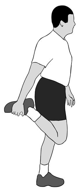
Flexibility exercises. Stretching your lower leg muscles may make your shins feel better.
Supportive shoes. Wearing shoes with good cushioning during daily activities will help reduce stress in your shins.
Orthotics. People who have flat feet or recurrent problems with shin splints may benefit from orthotics. Shoe inserts can help align and stabilize your foot and ankle, taking stress off of your lower leg. Orthotics can be custom-made for your foot, or purchased “off the shelf.”
Return to exercise. Shin splints usually resolve with rest and the simple treatments described above. Before returning to exercise, you should be pain-free for at least 2 weeks. Keep in mind that, when you return to exercise, it must be at a lower level of intensity. You should not be exercising as often as you did before, or for the same length of time.
Be sure to warm up and stretch thoroughly before you exercise. Increase training slowly. If you start to feel the same pain, stop exercising immediately. Use a cold pack and rest for a day or two. Return to training again at a lower level of intensity. Increase training even more slowly than before.
There are things you can do to prevent shin splints.
Wear a proper fitting athletic shoe. To get the right fit, determine the shape of your foot using the “wet test.” Step out of the shower onto a surface that will show your footprint, like a brown paper bag. If you have a flat foot, you will see an impression of your whole foot on the paper. If you have a high arch, you will only see the ball and heel of your foot. When shopping, look for athletic shoes that match your particular foot pattern.
In addition, make sure you wear shoes designed for your sport. Running long distances in court-type sneakers can contribute to shin splints.
Slowly build your fitness level. Increase the duration, intensity, and frequency of your exercise regimen gradually.
Cross train. Alternate jogging with lower impact sports like swimming or cycling.
Barefoot running. In recent years, barefoot running has gained in popularity. Many people claim it has helped to resolve shin splints. Some research indicates that barefoot running spreads out impact stresses among muscles, so that no area is overloaded. However, there is no clear evidence that barefoot running reduces the risk for any injury.
Like any significant change in your fitness regimen, a barefoot running program should be started very gradually. Begin with short distances to give your muscles and your feet time to adjust. Pushing too far, too fast can put you at risk for stress injuries. In addition, barefoot runners are at increased risk for cuts and bruises on their feet. Several brands of minimalist shoes with “toes” are available and these also require a slow working in period as your body adjusts to this different activity.
If your shin splints do not improve after rest and other methods described above, be sure to see a doctor to determine whether something else is causing your leg pain.
There are a several ways in which the kneecap can become unstable or dislocate. In many cases, the patella dislocates with very little force because of an abnormality in the structure of a child’s knee.
In children with normal knee structure, patellar dislocations are often the result of a direct blow or a fall onto the knee. This incidence is more common in high-impact sports, such as football.
Dislocations can occur without contact, as well. A common example is that of a right-handed baseball player who dislocates the right patella while swinging the bat. When the right foot is planted on the ground and the torso rotates during the swing, the patella lags behind, resulting in dislocation.
The symptoms associated with a patellar dislocation depend on how far out of place the patella has moved and how much damage occurred when it happened.
Some general symptoms your child may experience include:
If your child’s patella has slid back into place, you should see your doctor as soon as possible. If your child’s patella is still out of place, go to the emergency room.
During the examination, your doctor will ask you and your child about how the injury occurred and specific symptoms. Your doctor also will evaluate the range of motion, tenderness, and appearance of the knee.
Imaging tests can help your doctor diagnose patellar instability, as well as determine a treatment plan.
Sometimes a piece of bone or cartilage can dislodge or loosen when the patella dislocates. This can be seen on an x-ray or MRI scan.
Patellofemoral pain syndrome (PFPS) is a broad term used to describe pain in the front of the knee and around the patella, or kneecap. It is sometimes called “runner’s knee” or “jumper’s knee” because it is common in people who participate in sports—particularly females and young adults—but PFPS can occur in nonathletes, as well. The pain and stiffness caused by PFPS can make it difficult to climb stairs, kneel down, and perform other everyday activities.
Many things may contribute to the development of PFPS. Problems with the alignment of the kneecap and overuse from vigorous athletics or training are often significant factors.
Symptoms are often relieved with conservative treatment, such as changes in activity levels or a therapeutic exercise program.
Your knee is the largest joint in your body and one of the most complex. It is made up of the lower end of the femur (thighbone), the upper end of the tibia (shinbone), and the patella (kneecap).
Ligaments and tendons connect the femur to the bones of the lower leg. The four main ligaments in the knee attach to the bones and act like strong ropes to hold the bones together.
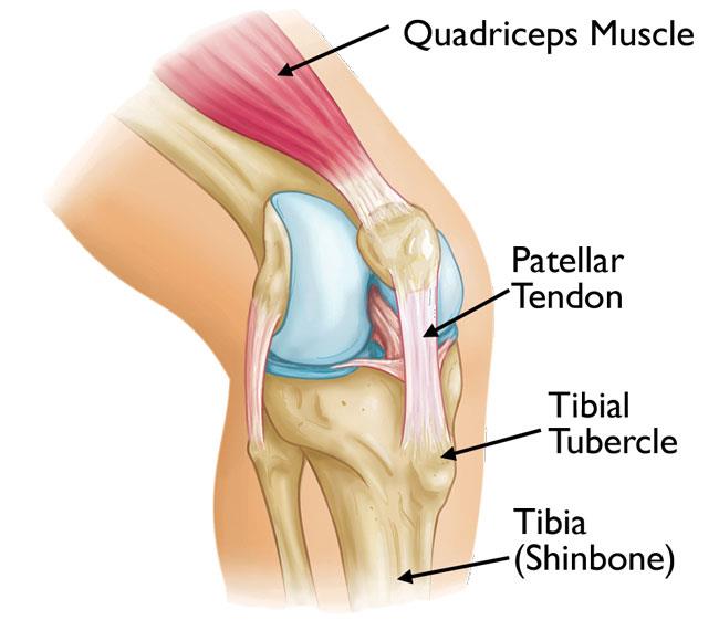
Muscles are connected to bones by tendons. The quadriceps tendon connects the muscles in the front of the thigh to the patella. Segments of the quadriceps tendon—called the patellar retinacula—attach to the tib
ia and help to stabilize the patella. Stretching from your patella to your tibia is the patellar tendon.
Several structures in the knee joint make movement easier. For example, the patella rests in a groove on the top of the femur called the trochlea. When you bend or straighten your knee, the patella moves back and forth inside this trochlear groove.
A slippery substance called articular cartilage covers the ends of the femur, trochlear groove, and the underside of the patella. Articular cartilage helps your bones glide smoothly against each other as you move your leg.
Also aiding in movement is the synovium—a thin lining of tissue that covers the surface of the joint. The synovium produces a small amount of fluid that lubricates the cartilage. In addition, just below the kneecap is a small pad of fat that cushions the kneecap and acts as a shock absorber.
Patellofemoral pain syndrome occurs when nerves sense pain in the soft tissues and bone around the kneecap. These soft tissues include the tendons, the fat pad beneath the patella, and the synovial tissue that lines the knee joint.
In some cases of patellofemoral pain, a condition called chondromalacia patella is present. Chondromalacia patella is the softening and breakdown of the articular cartilage on the underside of the kneecap. There are no nerves in articular cartilage—so damage to the cartilage itself cannot directly cause pain. It can, however, lead to inflammation of the synovium and pain in the underlying bone.
Patellofemoral pain syndrome can also be caused by abnormal tracking of the kneecap in the trochlear groove. In this condition, the patella is pushed out to one side of the groove when the knee is bent. This abnormality may cause increased pressure between the back of the patella and the trochlea, irritating soft tissues.
Factors that contribute to poor tracking of the kneecap include:
The most common symptom of PFPS is a dull, aching pain in the front of the knee. This pain—which usually begins gradually and is frequently activity-related—may be present in one or both knees. Other common symptoms include:
In many cases, patellofemoral pain will improve with simple home treatment.
Stop doing the activities that make your knee hurt until your pain is resolved. This may mean changing your training routine or switching to low-impact activities—such as riding a stationary bike, using an elliptical machine, or swimming—that will place less stress on your knee joint. If you are overweight, losing weight will also help to reduce pressure on your knee.
RICE stands for rest, ice, compression, and elevation.
Nonsteroidal anti-inflammatory drugs (NSAIDs) such as ibuprofen and naproxen can help reduce swelling and relieve pain.
If your pain persists or it becomes more difficult to move your knee, contact your doctor for a thorough evaluation.
During the physical examination, your doctor will discuss your general health and the symptoms you are experiencing. He or she will ask when your knee pain started and about the severity and nature of the pain (dull vs. sharp). Your doctor will also ask you what activities cause the pain to worsen.
To determine the exact location of the pain, your doctor may gently press and pull on the front of your knees and kneecaps. He or she may also ask you to squat, jump, or lunge during the exam in order to test your knee and core body strength.
To help diagnose the cause of your pain and to rule out any other physical problems, your doctor may also check:
Finally, your doctor may ask you to walk back and forth in order to examine your gait (the way you walk). He or she will look for problems with your gait that may be contributing to your knee pain.
X-rays. Usually, your doctor will be able to diagnose PFPS with just a physical examination. However, in most cases, he or she will also order an x-ray to rule out damage to the bones that make up the knee.
Magnetic resonance imaging (MRI) scans.This type of study provides clear images of the body’s soft tissues, such as ligaments, tendons, and muscles. Your doctor may order an MRI if, after a period of time, your symptoms do not improve with physical therapy and home exercise.
Medical treatment for PFPS is designed to relieve pain and restore range of motion and strength. In most cases, patellofemoral pain can be treated nonsurgically.
In addition to activity changes, the RICE method, and anti-inflammatory medication, your doctor may recommend the following:
Physical therapy. Specific exercises will help you improve range of motion, strength, and endurance. It is especially important to focus on strengthening and stretching your quadriceps since these muscles are the main stabilizers of your kneecap. Core exercises may also be recommended to strengthen the muscles in your abdomen and lower back.
Orthotics. Shoe inserts can help align and stabilize your foot and ankle, taking stress off of your lower leg. Orthotics can either be custom-made for your foot or purchased “off the shelf.”
Tibial tubercle transfer. In some cases, it may be necessary to realign the kneecap by moving the patellar tendon along with a portion of the tibial tubercle—the bony prominence on the tibia (shinbone).
A traditional open surgical incision is required for this procedure. The doctor partially or totally detaches the tibial tubercle so that the bone and the tendon can be moved toward the inner side of the knee. The piece of bone is then reattached to the tibia using screws. In most cases, this transfer allows for better tracking of the kneecap in the trochlear groove.
Patellofemoral pain syndrome is usually fully relieved with simple measures or physical therapy. It may recur, however, if you do not make adjustments to your training routine or activity level. It is essential to maintain appropriate conditioning of the muscles around the knee, particularly the quadriceps and the hamstrings.
There are additional steps that you can take to prevent recurrence of patellofemoral knee pain. They include:
Bursitis usually develops as the result of overuse or constant friction and stress on the bursa. Pes anserine bursitis is common in athletes, particularly runners. People with osteoarthritis of the knee are also susceptible.
Several factors can contribute to the development of pes anserine bursitis, including:
The symptoms of pes anserine bursitis include:
Your doctor will examine your knee and talk to you about your symptoms.
Symptoms of pes anserine bursitis may mimic those of a stress fracture, so an x-ray is usually required for diagnosis.
Athletes with pes anserine bursitis should take steps to modify their workout program so that the inflammation does not recur.
Other treatments include:
If your symptoms continue, your orthopaedic surgeon may recommend surgical removal of the bursa. This is typically performed as an outpatient (same-day) procedure.
If putting weight on your leg causes discomfort after the procedure, your doctor will recommend using crutches for a short time. Normal activities can typically be resumed within 3 weeks of the procedure.
The bones of children and adolescents possess a special area where the bone is growing called the growth plate. Growth plates are areas of cartilage located near the ends of bones. When a child is fully grown, the tibial tubercle covers the growth plate at the end of the tibia. The group of muscles in the front of the thigh (called the quadriceps) attaches to the tibial tubercle.
When a child is active, the quadriceps muscles pull on the patellar tendon which, in turn, pulls on the tibial tubercle. In some children, this repetitive traction on the tubercle leads to inflammation of the growth plate. The prominence, or bump, of the tibial tubercle may become very pronounced.
Painful symptoms are often brought on by running, jumping, and other sports-related activities. In some cases, both knees have symptoms, although one knee may be worse than the other.
During the appointment, your doctor will discuss your child’s symptoms and general health. He or she will conduct a thorough examination of the knee to determine the cause of the pain. This will include applying pressure to the tibial tubercle, which should be tender or painful for a child with Osgood-Schlatter disease. In addition, your doctor may also ask your child to walk, run, jump, or kneel to see if the movements bring on painful symptoms.
Your doctor may also order an x-ray image of your child’s knee to help confirm the diagnosis or rule out any other problems.
The knee is the largest and strongest joint in the body. It is made up of the lower end of the femur (thighbone), upper end of the tibia (shinbone), and the patella (kneecap). A smooth, slippery tissue called articular cartilage covers and protects the ends of the bones where they meet to form a joint.
The knee joint is filled with a clear fluid (synovial fluid) that acts as a lubricant to help reduce friction within the joint. Small fluid-filled sacs called bursa cushion the joint and help reduce friction between the muscles and other surrounding structures.
In adults, Baker’s cysts usually result from an injury or condition that causes swelling and inflammation inside the knee joint, such as:
In response to this inflammation, the knee produces excess synovial fluid, which travels behind the knee and accumulates in the popliteal bursa. The bursa then swells and bulges, forming a Baker’s cyst.
In younger patients, Baker’s cysts often have no known cause.
Some Baker’s cysts cause no symptoms and are only discovered incidentally during a physical exam or when an MRI scan is performed for some other reason. When symptoms do occur, they may include:
If the cyst becomes very large, it can interfere with blood flow in the veins of your leg. This can cause pain, swelling, weakness, or even numbness if there is nerve compression. Very rarely, the cyst can even burst.
Sometimes the symptoms of a cyst may resemble those of a blood clot or deep vein thrombosis, a much more serious problem. If you have increasing pain and swelling in your calf, it is important to seek medical care right away to rule out a blood clot.
Your doctor will take a full medical history and ask you to describe your symptoms. He or she will want to know if you have had a previous knee injury.
Your doctor will then perform a careful examination of your affected knee, comparing it to your “normal” knee. During the exam, he or she will look for:
Your doctor will also palpate (feel) the back of your knee where the cyst is located. Often, a cyst will become firm when the knee is fully extended and soft when the knee is bent.
Imaging tests may be ordered to help confirm the diagnosis and provide more information about your condition.
X-rays. X-rays provide images of dense structures, such as bone. Although a cyst cannot be seen on an x-ray, one may be ordered so that your doctor can look for narrowing of the joint space and other signs of arthritis in the joint.
Ultrasound. This test uses sound waves to create images of structures inside the body. An ultrasound will help your doctor see the lump behind your knee in greater detail and determine whether it is solid or filled with fluid.
Magnetic resonance imaging (MRI) scans. These tests produce clear pictures of the body’s soft tissues. Your doctor may order an MRI scan to learn more about your cyst and to look for a meniscus tear or another underlying condition.
The camera attached to the arthroscope displays the image of the joint on a video monitor, allowing the surgeon to look, for example, throughout the knee. This lets the surgeon see the cartilage, ligaments, and under the kneecap. The surgeon can determine the amount or type of injury and then repair or correct the problem, if it is necessary.
Your bones, cartilage, ligaments, muscles, and tendons can all be damaged by disease and injury. To diagnose your condition, your doctor will take a thorough medical history, perform a physical examination and order imaging studies — usually x-rays. For some conditions, an additional imaging study — such as a magnetic resonance imaging (MRI) scan or a computerized tomography (CT) scan — may also be needed. After making a diagnosis, your doctor will determine the most appropriate treatment choice for your condition.
Conditions that are commonly treated with arthroscopic procedures include:
Some problems associated with arthritis can also be treated arthroscopically.
The procedures below are performed either with arthroscopy or with a combination of arthroscopic and open surgery:
Although the inside of nearly all joints can be viewed with an arthroscope, six joints are most frequently examined with this instrument. These are the knee, shoulder, elbow, ankle, hip and wrist. As advances are made in fiberoptic technology and new techniques are developed by orthopaedic surgeons, other joints may be treated more frequently in the future.
Arthroscopic surgery, although much easier in terms of recovery than open surgery, still requires the use of anesthetics and the special equipment in a hospital operating room or outpatient surgical suite. You will be given a general, spinal, or a local anesthetic, depending on the joint or suspected problem.
A small incision (about the size of a buttonhole) will be made to insert the arthroscope. Several other incisions may be made to see other parts of the joint or insert other instruments.
Corrective surgery is performed with specially designed instruments that are inserted into the joint through accessory incisions. Originally, arthroscopy was simply a diagnostic tool used for planning standard open surgery. However, with the development of better instrumentation and surgical techniques, many conditions can now be treated with arthroscopic techniques.
After arthroscopic surgery, the small incisions will be covered with a dressing. You will be moved from the operating room to a recovery room. Many patients need little or no pain medication.
Before being discharged, you will be given instructions on caring for your incisions, what activities to avoid, and which exercises you should do to aid your recovery. During the follow-up visit, the surgeon will inspect your incisions; remove sutures, if present; and discuss your rehabilitation program.
The amount of surgery required and recovery time will depend on the complexity of your problem. Occasionally, during arthroscopy, the surgeon may discover that the injury or disease cannot be treated adequately with arthroscopy alone. More extensive open surgery may be performed while you are still anesthetized, or at a later date after you have discussed the findings with your surgeon.
Although uncommon, complications do occur occasionally during or after arthroscopy. Infection, blood clots of a vein (also called deep vein thrombosis or DVT), excessive swelling or bleeding, damage to blood vessels or nerves, and instrument breakage are the most common complications; however, these occur in far less than 1 percent of all arthroscopic procedures.
Although arthroscopic surgery has received a lot of public attention because it is often used to treat well-known athletes, it is an extremely valuable tool for all orthopaedic patients and is generally easier on the patient than open surgery. Most patients have their arthroscopic surgery as outpatients and are home several hours after the surgery.
The small puncture wounds take several days to heal. The operative dressing can usually be removed the morning after surgery and adhesive strips can be applied to cover the small healing incisions.
Although the puncture wounds are small and pain in the joint that underwent arthroscopy is minimal, it takes several weeks for the joint to maximally recover. A specific activity and rehabilitation program may be suggested to speed your recover and protect future joint function.
It is not unusual for patients to go back to work or school or resume daily activities within a few days. Athletes and others who are in good physical condition may, in some cases, return to athletic activities within a few weeks. Remember, though, that people who have arthroscopy can have many different diagnoses and preexisting conditions, so each patient’s arthroscopic surgery is unique to that person. Recovery time will reflect that individuality.
The bone structure of the knee joint is formed by the femur, the tibia, and the patella. The ACL is one of the four main ligaments within the knee that connect the femur to the tibia.
The knee is essentially a hinged joint that is held together by the medial collateral (MCL), lateral collateral (LCL), anterior cruciate (ACL) and posterior cruciate (PCL) ligaments. The ACL runs diagonally in the middle of the knee, preventing the tibia from sliding out in front of the femur, as well as providing rotational stability to the knee.
The weight-bearing surface of the knee is covered by a layer of articular cartilage. On either side of the joint, between the cartilage surfaces of the femur and tibia, are the medial meniscus and lateral meniscus. The menisci act as shock absorbers and work with the cartilage to reduce the stresses between the tibia and the femur.
The anterior cruciate ligament (ACL) is one of the most commonly injured ligaments of the knee. In general, the incidence of ACL injury is higher in people who participate in high-risk sports, such as basketball, football, skiing, and soccer.
Approximately half of ACL injuries occur in combination with damage to the meniscus, articular cartilage, or other ligaments. Additionally, patients may have bruises of the bone beneath the cartilage surface. These may be seen on a magnetic resonance imaging (MRI) scan and may indicate injury to the overlying articular cartilage.
It is estimated that the majority of ACL injuries occur through non-contact mechanisms, while a smaller percent result from direct contact with another player or object.
The mechanism of injury is often associated with deceleration coupled with cutting, pivoting or sidestepping maneuvers, awkward landings or “out of control” play.
Several studies have shown that female athletes have a higher incidence of ACL injury than male athletes in certain sports. It has been proposed that this is due to differences in physical conditioning, muscular strength, and neuromuscular control. Other hypothesized causes of this gender-related difference in ACL injury rates include pelvis and lower extremity (leg) alignment, increased ligamentous laxity, and the effects of estrogen on ligament properties.
Immediately after the injury, patients usually experience pain and swelling and the knee feels unstable. Within a few hours after a new ACL injury, patients often have a large amount of knee swelling, a loss of full range of motion, pain or tenderness along the joint line and discomfort while walking.
When a patient with an ACL injury is initially seen for evaluation in the clinic, the doctor may order x-rays to look for any possible fractures. He or she may also order a magnetic resonance imaging (MRI) scan to evaluate the ACL and to check for evidence of injury to other knee ligaments, meniscus cartilage, or articular cartilage.
In addition to performing special tests for identifying meniscus tears and injury to other ligaments of the knee, the physician will often perform the Lachman’s test to see if the ACL is intact.
If the ACL is torn, the examiner will feel increased forward (upward or anterior) movement of the tibia in relation to the femur (especially when compared to the normal leg) and a soft, mushy endpoint (because the ACL is torn) when this movement ends.
What happens naturally with an ACL injury without surgical intervention varies from patient to patient and depends on the patient’s activity level, degree of injury and instability symptoms.
The prognosis for a partially torn ACL is often favorable, with the recovery and rehabilitation period usually at least 3 months. However, some patients with partial ACL tears may still have instability symptoms. Close clinical follow-up and a complete course of physical therapy helps identify those patients with unstable knees due to partial ACL tears.
Complete ACL ruptures have a much less favorable outcome without surgical intervention. After a complete ACL tear, some patients are unable to participate in cutting or pivoting-type sports, while others have instability during even normal activities, such as walking. There are some rare individuals who can participate in sports without any symptoms of instability. This variability is related to the severity of the original knee injury, as well as the physical demands of the patient.
About half of ACL injuries occur in combination with damage to the meniscus, articular cartilage or other ligaments. Secondary damage may occur in patients who have repeated episodes of instability due to ACL injury. With chronic instability, a large majority of patients will have meniscus damage when reassessed 10 or more years after the initial injury. Similarly, the prevalence of articular cartilage lesions increases in patients who have a 10-year-old ACL deficiency.
In nonsurgical treatm
ent, progressive physical therapy and rehabilitation can restore the knee to a condition close to its pre-injury state and educate the patient on how to prevent instability. This may be supplemented with the use of a hinged knee brace. However, many people who choose not to have surgery may experience secondary injury to the knee due to repetitive instability episodes.
Surgical treatment is usually advised in dealing with combined injuries (ACL tears in combination with other injuries in the knee). However, deciding against surgery is reasonable for select patients. Nonsurgical management of isolated ACL tears is likely to be successful or may be indicated in patients:
ACL tears are not usually repaired using suture to sew it back together, because repaired ACLs have generally been shown to fail over time. Therefore, the torn ACL is generally replaced by a substitute graft made of tendon.
Active adult patients involved in sports or jobs that require pivoting, turning or hard-cutting as well as heavy manual work are encouraged to consider surgical treatment. This includes older patients who have previously been excluded from consideration for ACL surgery. Activity, not age, should determine if surgical intervention should be considered.
In young children or adolescents with ACL tears, early ACL reconstruction creates a possible risk of growth plate injury, leading to bone growth problems. The surgeon can delay ACL surgery until the child is closer to skeletal maturity or the surgeon may modify the ACL surgery technique to decrease the risk of growth plate injury.
A patient with a torn ACL and significant functional instability has a high risk of developing secondary knee damage and should therefore consider ACL reconstruction.
It is common to see ACL injuries combined with damage to the menisci, articular cartilage, collateral ligaments, joint capsule, or a combination of the above. The “unhappy triad,” frequently seen in football players and skiers, consists of injuries to the ACL, the MCL, and the medial meniscus.
In cases of combined injuries, surgical treatment may be warranted and generally produces better outcomes. As many as half of meniscus tears may be repairable and may heal better if the repair is done in combination with the ACL reconstruction.
Patellar tendon autograft. The middle third of the patellar tendon of the patient, along with a bone plug from the shin and the kneecap is used in the patellar tendon autograft. Occasionally referred to by some surgeons as the “gold standard” for ACL reconstruction, it is often recommended for high-demand athletes and patients whose jobs do not require a significant amount of kneeling.
In studies comparing outcomes of patellar tendon and hamstring autograft ACL reconstruction, the rate of graft failure was lower in the patellar tendon group. In addition, most studies show equal or better outcomes in terms of postoperative tests for knee laxity (Lachman’s, anterior drawer and instrumented tests) when this graft is compared to others. However, patellar tendon autografts have a greater incidence of postoperative patellofemoral pain (pain behind the kneecap) complaints and other problems.
The pitfalls of the patellar tendon autograft are:
Hamstring tendon autograft. The semitendinosus hamstring tendon on the inner side of the knee is used in creating the hamstring tendon autograft for ACL reconstruction. Some surgeons use an additional tendon, the gracilis, which is attached below the knee in the same area. This creates a two- or four-strand tendon graft. Hamstring graft proponents claim there are fewer problems associated with harvesting of the graft compared to the patellar tendon autograft including:
The graft function may be limited by the strength and type of fixation in the bone tunnels, as the graft does not have bone plugs. There have been conflicting results in research studies as to whether hamstring grafts are slightly more susceptible to graft elongation (stretching), which may lead to increased laxity during objective testing. Recently, some studies have demonstrated decreased hamstring strength in patients after surgery.
There are some indications that patients who have intrinsic ligamentous laxity and knee hyperextension of 10 degrees or more may have increased risk of postoperative hamstring graft laxity on clinical exam. Therefore, some clinicians recommend the use of patellar tendon autografts in these hypermobile patients.
Additionally, since the medial hamstrings often provide dynamic support against valgus stress and instability, some surgeons feel that chronic or residual medial collateral ligament laxity (grade 2 or more) at the time of ACL reconstruction may be a contraindication for use of the patient’s own semitendinosus and gracilis tendons as an ACL graft.
Quadriceps tendon autograft. The quadriceps tendon autograft is often used for patients who have already failed ACL reconstruction. The middle third of the patient’s quadriceps tendon and a bone plug from the upper end of the knee cap are used. This yields a larger graft for taller and heavier patients. Because there is a bone plug on one side only, the fixation is not as solid as for the patellar tendon graft. There is a high association with postoperative anterior knee pain and a low risk of patella fracture. Patients may find the incision is not cosmetically appealing.
Allografts. Allografts are grafts taken from cadavers and are becoming increasingly popular. These grafts are also used for patients who have failed ACL reconstruction before and in surgery to repair or reconstruct more than one knee ligament. Advantages of using allograft tissue include elimination of pain caused by obtaining the graft from the patient, decreased surgery time and smaller incisions. The patellar tendon allograft allows for strong bony fixation in the tibial and femoral bone tunnels with screws.
However, allografts are associated with a risk of infection, including viral transmission (HIV and Hepatitis C), despite careful screening and processing. Several deaths linked to bacterial infection from allograft tissue (due to improper procurement and sterilization techniques) have led to improvements in allograft tissue testing and processing techniques. There have also been conflicting results in research studies as to whether allografts are slightly more susceptible to graft elongation (stretching), which may lead to increased laxity during testing.
Some published literature may point to a higher failure rate with the use of allografts for ACL reconstruction. Higher failure rates for allografts have been reported in young, active patients returning to high-demand sporting activities after ACL reconstruction, compared with autografts.
The reason for this higher failure rate is unclear. It could be due to graft material properties (sterilization processes used, graft donor age, storage of the graft). It could possibly be due to an ill-advised earlier return to sport by the athlete because of a faster perceived physiologic recovery, when the graft is not biologically ready to be loaded and stressed during sporting activities. Further research in this area is indicated and is ongoing.
Before any surgical treatment, the patient is usually sent to physical therapy. Patients who have a stiff, swollen knee lacking full range of motion at the time of ACL surgery may have significant problems regaining motion after surgery. It usually takes three or more weeks from the time of injury to achieve full range of motion. It is also recommended that some ligament injuries be braced and allowed to heal prior to ACL surgery.
The patient, the surgeon, and the anesthesiologist select the anesthesia used for surgery. Patients may benefit from an anesthetic block of the nerves of the leg to decrease postoperative pain.
The surgery usually begins with an examination of the patient’s knee while the patient is relaxed due the effects of anesthesia. This final examination is used to verify that the ACL is torn and also to check for looseness of other knee ligaments that may need to be repaired during surgery or addressed postoperatively.
If the physical exam strongly suggests the ACL is torn, the selected tendon is harvested (for an autograft) or thawed (for an allograft) and the graft is prepared to the correct size for the patient.
After the graft has been prepared, the surgeon places an arthroscope into the joint. Small (one-centimeter) incisions called portals are made in the front of the knee to insert the arthroscope and instruments and the surgeon examines the condition of the knee. Meniscus and cartilage injuries are trimmed or repaired and the torn ACL stump is then removed.
In the most common ACL reconstruction technique, bone tunnels are drilled into the tibia and the femur to place the ACL graft in almost the same position as the torn ACL. A long needle is then passed through the tunnel of the tibia, up through the femoral tunnel, and then out through the skin of the thigh. The sutures of the graft are placed through the eye of the needle and the graft is pulled into position up through the tibial tunnel and then up into the femoral tunnel. The graft is held under tension as it is fixed in place using interference screws, spiked washers, posts, or staples. The devices used to hold the graft in place are generally not removed.
Variations on this surgical technique include the “two-incision,” “over-the-top,” and “double-bundle” types of ACL reconstructions, which may be used because of the preference of the surgeon or special circumstances (revision ACL reconstruction, open growth plates).
Before the surgery is complete, the surgeon will probe the graft to make sure it has good tension, verify that the knee has full range of motion and perform tests such as the Lachman’s test to assess graft stability. The skin is closed and dressings (and perhaps a postoperative brace and cold therapy device, depending on surgeon preference) are applied. The patient will usually go home on the same day of the surgery.
After surgery, you will feel some pain. This is a natural part of the healing process. Your doctor and nurses will work to reduce your pain, which can help you recover from surgery faster.
Medications are often prescribed for short-term pain relief after surgery. Many types of medicines are available to help manage pain, including opioids, non-steroidal anti-inflammatory drugs (NSAIDs), and local anesthetics. Your doctor may use a combination of these medications to improve pain relief, as well as minimize the need for opioids.
Be aware that although opioids help relieve pain after surgery, they are a narcotic and can be addictive. Opioid dependency and overdose has become a critical public health issue in the U.S. It is important to use opioids only as directed by your doctor. As soon as your pain begins to improve, stop taking opioids. Talk to your doctor if your pain has not begun to improve within a few days of your surgery.
Physical therapy is a crucial part of successful ACL surgery, with exercises beginning immediately after the surgery. Much of the success of ACL reconstructive surgery depends on the patient’s dedication to rigorous physical therapy. With new surgical techniques and stronger graft fixation, current physical therapy uses an accelerated course of rehabilitation.
Postoperative Course. In the first 10 to 14 days after surgery, the wound is kept clean and dry, and early emphasis is placed on regaining the ability to fully straighten the knee and restore quadriceps control.
The knee is iced regularly to reduce swelling and pain. The surgeon may dictate the use of a postoperative brace and the use of a machine to move the knee through its range of motion. Weight-bearing status (use of crutches to keep some or all of the patient’s weight off of the surgical leg) is also determined by physician preference, as well as other injuries addressed at the time of surgery.
Rehabilitation. The goals for rehabilitation of ACL reconstruction include reducing knee swelling, maintaining mobility of the kneecap to prevent anterior knee pain problems, regaining full range of motion of the knee, as well as strengthening the quadriceps and hamstring muscles.
The patient may return to sports when there is no longer pain or swelling, when full knee range of motion has been achieved, and when muscle strength, endurance and functional use of the leg have been fully restored.
The patient’s sense of balance and control of the leg must also be restored through exercises designed to improve neuromuscular control. This usually takes 4 to 6 months. The use of a functional brace when returning to sports is ideally not needed after a successful ACL reconstruction, but some patients may feel a greater sense of security by wearing one.
Infection. The incidence of infection after arthroscopic ACL reconstruction is very low. There have also been reported deaths linked to bacterial infection from allograft tissue due to improper procurement and sterilization techniques.
Viral transmission. Allografts specifically are associated with risk of viral transmission, including HIV and Hepatitis C, despite careful screening and processing. The chance of obtaining a bone allograft from an HIV-infected donor is calculated to be less than 1 in a million.
Bleeding, numbness. Rare risks include bleeding from acute injury to the popliteal artery, and weakness or paralysis of the leg or foot. It is not uncommon to have numbness of the outer part of the upper leg next to the incision, which may be temporary or permanent.
Blood clot. Although rare, blood clot in the veins of the calf or thigh is a potentially life-threatening complication. A blood clot may break off in the bloodstream and travel to the lungs, causing pulmonary embolism or to the brain, causing stroke.
Instability. Recurrent instability due to rupture or stretching of the reconstructed ligament or poor surgical technique is possible.
Stiffness. Knee stiffness or loss of motion has been reported by some patients after surgery.
Extensor mechanism failure. Rupture of the patellar tendon (patellar tendon autograft) or patella fracture (patellar tendon or quadriceps tendon autografts) may occur due to weakening at the site of graft harvest.
Growth plate injury. In young children or adolescents with ACL tears, early ACL reconstruction creates a possible risk of growth plate injury, leading to bone growth problems. The ACL surgery can be delayed until the child is closer to reaching skeletal maturity. Alternatively, the surgeon may be able to modify the technique of ACL reconstruction to decrease the risk of growth plate injury.
Kneecap pain. Postoperative anterior knee pain is especially common after patellar tendon autograft ACL reconstruction. The incidence of pain behind the kneecap varies greatly in studies, whereas the incidence of kneeling pain is often higher after patellar tendon autograft ACL reconstruction.
Partial meniscectomy is a surgical procedure to remove the torn portion of the meniscus from the knee joint. Meniscus is the C-shaped cartilage located in the knee that lubricates the knee joint, acts as shock-absorber, and controls the flexion and extension of joint.
Meniscal tears can occur at any age, but are more common in athletes playing contact sports. These tears are usually caused by twisting motion or over flexing of the knee joint. Athletes who play sports, such as football, tennis and basketball are at a higher risk of developing meniscal tears.
You may have pain over inner and outer side of the knee, swelling, stiffness of knee, restricted movement of the knee, and difficulty in straightening your knee. If the conservative treatment such as pain medications, rest, physical therapy, and use of knee immobilizers fails to relieve pain, then surgery may be recommended. Surgical treatment options depend on the location, length, and pattern of the tear.
There are two surgical procedures for meniscal tears which includes total and partial meniscectomy. In total meniscectomy, the entire meniscus is removed, but in partial meniscectomy your surgeon will only remove the torn meniscus. Total meniscectomy will help in relieving symptoms, but because the entire meniscus is removed; the cushioning and stability between the joints will be lost. Hence partial meniscectomy is considered.
Partial meniscectomy helps in restoring or maintaining knee stability and offers faster and complete recovery. After surgery rehabilitation exercises may help to restore knee mobility, strength and to improve range of motion.
Possible risks and complications of partial meniscectomy include infection, bleeding, and injury to blood vessels or nerves.
The knee is the largest joint of the body and is stabilized by a set of ligaments. In the knee, there are four primary ligaments viz. anterior cruciate ligament (ACL), posterior cruciate ligament (PCL), medial collateral ligament (MCL) and lateral collateral ligament.
Lateral collateral ligament may tear due to trauma, sports injuries, or direct blow on the knee. Torn LCL may result in pain, swelling and even instability of the knee.
LCL injuries and torn LCL can be diagnosed through physical examination and by employing imaging techniques such as X-rays or MRI scan.
The treatment of the torn LCL include non-surgical interventions such as rest, ice, elevation, bracing and physical therapy to help reduce swelling, and regain activity as well as strength and flexibility of the knee. Surgery is recommended if non-surgical interventions fail to provide much relief. Surgical interventions include repair and reconstruction of the torn ligament. Based on the severity and location of the injury, repair or reconstruction of the LCL is recommended. In case the ligament is torn from the upper or lower ends of attachment, then repair of the LCL is done with sutures or staples. If the ligament is torn in the middle or if the injury is older than 3 weeks, LCL reconstruction is recommended.
LCL reconstruction involves replacement of the torn ligament with healthy strong tissue or graft. The tissue or graft can be taken either from the tissue bank (called allograft) or from the patient’s body (called autograft). The type of graft used, depends upon the condition of the patient and choice of your surgeon. Hamstring tendons are commonly used as autograft, as removal of such tendons does not affect the strength of the legs and effectively stabilizes the knee. A small incision is made on the lateral side of the knee to perform the LCL reconstruction. The procedure is done through an open incision and not arthroscopically. The thighbone and fibula bones are drilled precisely and accurately with specialized instruments to form tunnels. The ends of the tendon graft are passed through tunnels and are fixed by using screws, metal staples or large sutures. The knee undergone LCL reconstruction surgery is braced for 6-8 weeks.
The common Post-operative instructions for LCL reconstruction are:
Some of the possible risks and complication associated with LCL reconstruction include:
Three bones meet to form your knee joint: your thighbone (femur), shinbone (tibia), and kneecap (patella). Your kneecap sits in front of the joint to provide some protection.
Bones are connected to other bones by ligaments. There are four primary ligaments in your knee. They act like strong ropes to hold the bones together and keep your knee stable.
These are found inside your knee joint. They cross each other to form an “X” with the anterior cruciate ligament in front and the posterior cruciate ligament in back. The cruciate ligaments control the back and forth motion of your knee.
These are found on the sides of your knee. The medial or “inside” collateral ligament (MCL) connects the femur to the tibia. The lateral or “outside” collateral ligament (LCL) connects the femur to the smaller bone in the lower leg (fibula). The collateral ligaments control the sideways motion of your knee and brace it against unusual movement.
Because the knee joint relies just on these ligaments and surrounding muscles for stability, it is easily injured. Any direct contact to the knee or hard muscle contraction — such as changing direction rapidly while running — can injure a knee ligament.
Injured ligaments are considered “sprains” and are graded on a severity scale.
Grade 1 Sprains. The ligament is mildly damaged in a Grade 1 Sprain. It has been slightly stretched, but is still able to help keep the knee joint stable.
Grade 2 Sprains. A Grade 2 Sprain stretches the ligament to the point where it becomes loose. This is often referred to as a partial tear of the ligament.
Grade 3 Sprains. This type of sprain is most commonly referred to as a complete tear of the ligament. The ligament has been split into two pieces, and the knee joint is unstable..
The MCL is injured more often than the LCL. Due to the more complex anatomy of the outside of the knee, if you injure your LCL, you usually injure other structures in the joint, as well.
Injuries to the collateral ligaments are usually caused by a force that pushes the knee sideways. These are often contact injuries, but not always.
Medial collateral ligament tears often occur as a result of a direct blow to the outside of the knee. This pushes the knee inwards (toward the other knee).
Blows to the inside of the knee that push the knee outwards may injure the lateral collateral ligament.
During your first visit, your doctor will talk to you about your symptoms and medical history.
During the physical examination, your doctor will check all the structures of your injured knee, and compare them to your non-injured knee. Most ligament injuries can be diagnosed with a thorough physical examination of the knee.
Other tests which may help your doctor confirm your diagnosis include:
X-rays. Although they will not show any injury to your collateral ligaments, x-rays can show whether the injury is associated with a broken bone.
Magnetic resonance imaging (MRI) scan. This study creates better images of soft tissues like the collateral ligaments.
Injuries to the MCL rarely require surgery. If you have injured just your LCL, treatment is similar to an MCL sprain. But if your LCL injury involves other structures in your knee, your treatment will address those, as well.
Ice. Icing your injury is important in the healing process. The proper way to ice an injury is to use crushed ice directly to the injured area for 15 to 20 minutes at a time, with at least 1 hour between icing sessions. Chemical cold products (“blue” ice) should not be placed directly on the skin and are not as effective.
Bracing. Your knee must be protected from the same sideways force that caused the injury. You may need to change your daily activities to avoid risky movements. Your doctor may recommend a brace to protect the injured ligament from stress. To further protect your knee, you may be given crutches to keep you from putting weight on your leg.
Physical therapy. Your doctor may suggest strengthening exercises. Specific exercises will restore function to your knee and strengthen the leg muscles that support it.
Most isolated collateral ligament injuries can be successfully treated without surgery. If the collateral ligament is torn in such a way that it cannot heal or is associated with other ligament injuries, your doctor may suggest surgery to repair it.
Once your range of motion returns and you can walk without a limp, your doctor may allow functional progression. This is a gradual, progressive return to sports activities.
For example, if you play soccer, your functional progression may start as a light jog. Then you progress to a sprint, and eventually to full running and kicking the ball.
Your doctor may suggest a knee brace during sports activities, depending on the severity of your sprain.
Medial patellofemoral ligament reconstruction is a surgical procedure indicated in patients with more severe patellar instability. Medial patellofemoral ligament is a band of tissue that extends from the femoral medial epicondyle to the superior aspect of the patella. Medial patellofemoral ligament is the major ligament which stabilizes the patella and helps in preventing patellar subluxation (partial dislocation) or dislocation. This ligament can rupture or get damaged when there is patellar lateral dislocation. Dislocation can be caused by direct blow to the knee, twisting injury to the lower leg, strong muscle contraction, or because of a congenital abnormality such as shallow or malformed joint surfaces.
Medial patellofemoral ligament reconstruction using autogenous tissue grafts is done by following the basic principles of ligament reconstruction such as:
The surgical procedure of medial patellofemoral ligament reconstruction involves the following steps:
Graft Selection and Harvest:
Your surgeon will make a 4-6 cm skin incision over your knee, at the midpoint between the medial epicondyle and the medial aspect of the patella (kneecap). The underlying subcutaneous fat and fascia are cut apart to expose the adductor tendon. The tendon is then stripped using a tendon stripper and its free end is sutured. The diameter of the tendon graft is measured using a sizer.
Alternatively, a graft can be harvested from the quadriceps tendon.
Location of the Femoral Isometric Point:
The graft should be placed isometrically to prevent it from overstretching and causing failure during joint movements. A transverse hole measuring 2.5 mm is made through the patella. Then a small incision is made over the lateral side of the patella and a strand of Vicryl suture material is inserted through the hole. Over this strand, a 2.5 mm Kirschner wire (K-wire) is passed and then inserted into the bone besides the medial epicondyle.
An instrument called pneumatic isometer is inserted into the hole made in the patella and the Vicryl isometric measurement suture material is also passed along. The knee is taken through its full range of motion and any changes happening in the length between the medial epicondylar K-wire and the medial aspect of the patella is recorded on the isometer. The position of the K-wire will be adjusted until no deviations are read on the isometer during full range of motion. Once the isometric point is identified, a tunnel is drilled starting from the insertion of the adductor tendon uptil the isometric point is reached. The graft is pulled through this tunnel, then exits at the medial condyle and again passed through another tunnel that is made through the patella.
Correct Tension:
The tension is set in the graft with your knee flexed up to 90o and the tension should be appropriate enough to control lateral excursion.
Secure Fixation:
After bringing the tendon graft from the medial to the lateral side through the bone tunnel, it turned onto the front surface of the patella where it is sutured.
Avoid Condylar Rubbing and Impingement:
After graft fixation, the range of motion is checked to make sure there are no restrictions in patellar or knee movements. The graft should not impinge or rub against the medial femoral condyle. If it is detected, the graft is replaced into proper position.
A knee brace should be used during walking in the first 3-6 weeks after surgery. Avoid climbing stairs, squatting and stretching your leg until there is adequate healing of the tendon. Rehabilitation exercises, continuous passive motion and active exercises will be recommended.
Tibial tubercle osteotomy is a surgical procedure which is performed along with other procedures to treat patellar instability, patellofemoral pain, and osteoarthritis. Tibial tubercle transfer technique involves realignment of the tibial tubercle (a bump in the front of the shinbone) such that the kneecap (patella) traverses in the center of the femoral groove. The patellar maltracking is corrected by moving the tibial tubercle medially, towards the inside portion of the leg. This removes the load off the painful portions of the kneecap and reduces the pain.
Surgical treatment is indicated when physical therapy and other nonsurgical methods have failed and there is history of multiple knee dislocations.
The procedure is performed under general anesthesia and you will be completely unaware of the surgery until you wake up in the recovery room. At first, knee arthroscopy will be performed to inspect the inside portions of the knee joint. It involves small incisions or portals through which small instruments are passed and a video camera is used to visualize the anatomy of the knee joint, evaluate patella cartilage and assess patella tracking.
Tibial tubercle osteotomy and transfer is done through an incision made in the front of your leg just below the patella. In osteotomy procedure, a periosteal incision of 8-10 cm length is made at 1cm medial to the tibial tubercle. With the help of an oscillating saw, a cut is made medial to the tuberosity and a distal cut is also made. The tapered design of the distal cut avoids the risk of tibial fracture. Similarly, a proximal cut is made using appropriate instruments such as curved osteotome or reciprocating saw. Then an osteotomy through the bone cortex is performed without cutting off the lateral periosteum. The lateral periosteum serves as a point of attachment for the osteotomy segment. By doing this, a tibial tubercle segment which is more than 2 cm in width, more than 1 cm in thickness and 8-10 cm length can be obtained. It should include all portions of insertion of the patellar tendon. The segment from the tibia is then levered using osteotome to provide access to the medullary canal of the tibia.
The osteotomy segment is then moved under direct vision into a position that assures proper tracking of the patella. The tracking pattern can be confirmed arthroscopically. The mobilized bone is then fixed into its new place using screws, which can be removed later if they cause irritation.
You may have minimal to moderate knee discomfort for several days or weeks after the surgery. Oral pain medications will be prescribed that helps control your pain. Keep the operated leg elevated and apply ice bag over the area for 20 minutes. This decrease swelling as well as pain. You will have a leg brace which may be removed only while sitting with your leg elevated and when using the continuous passive motion (CPM) unit. Physical therapy exercises should be done as it helps in regaining mobility. Eat healthy food and drink plenty of water.
This is a quite safe procedure and provides excellent access and surgical exposure during a difficult primary or revision total knee arthroplasty. Risks following tibial tubercle osteotomy surgery are rare but may include compartment syndrome, deep vein thrombosis, infections and delayed bone healing.