Simply defined, tendinitis is inflammation of a tendon. Inflammation is the body’s natural response to injury or disease, and often causes swelling, pain, or irritation.
There are two types of Achilles tendinitis, based upon which part of the tendon is inflamed.
In noninsertional Achilles tendinitis, fibers in the middle portion of the tendon have begun to break down with tiny tears (degenerate), swell, and thicken.
Tendinitis of the middle portion of the tendon more commonly affects younger, active people.
Insertional Achilles tendinitis involves the lower portion of the heel, where the tendon attaches (inserts) to the heel bone.
In both noninsertional and insertional Achilles tendinitis, damaged tendon fibers may also calcify (harden). Bone spurs (extra bone growth) often form with insertional Achilles tendinitis.
Tendinitis that affects the insertion of the tendon can occur at any time, even in patients who are not active. More often than not, however, it comes from years of overuse (long distance runners, sprinters).
Achilles tendinitis is typically not related to a specific injury. The problem results from repetitive stress to the tendon. This often happens when we push our bodies to do too much, too soon, but other factors can make it more likely to develop tendinitis, including:
Females in any sport can develop one or more components of the triad. At greatest risk are those in sports that reward being thin for appearance (such as figure skating or gymnastics) or improved performance (such as distance running or rowing).
Fashion trends and advertising often encourage women to try to reach unhealthy weight levels. Some female athletes suffer low self-esteem or depression, and may focus on weight loss because they think they are heavier than they actually are. Others feel pressure to lose weight from athletic coaches or parents.
Female athletes should consider these questions:
If the answer to any of these questions is yes, you may be at risk for developing abnormal patterns of eating (disordered eating), which can lead to menstrual dysfunction and early osteoporosis.
Although they usually do not realize or admit that they are ill, people with disordered eating have serious and complex disturbances in eating behaviors. They are preoccupied with body shape and weight and have poor nutritional habits.
Disordered eating can take many forms. Some people starve themselves (anorexia nervosa) or engage in cycles of overeating and purging (bulimia).
Others severely restrict the amount of food they eat, fast for prolonged periods of time or misuse diet pills, diuretics, or laxatives. People with disordered eating may also exercise excessively to keep their weight down.
Females are more likely than males to have disordered eating. The illness can cause many problems, including dehydration, muscle fatigue and weakness, an erratic heartbeat, kidney damage, and other serious conditions. Not taking in enough calcium can lead to bone loss. It is especially bad to lose bone when you are a child or teenager because that is when your body should be building bone. Hormone imbalances can lead to more bone loss through menstrual dysfunction.
Missing three or more periods in a row is cause for concern. With normal menstruation, the body produces estrogen, a hormone that helps to keep bones strong. Without a menstrual cycle (amenorrhea), the level of estrogen may be lowered, causing a loss of bone density and strength (premature osteoporosis).
If this happens during youth, it may become a serious problem later in life when the natural process of bone mineral loss begins after menopause. Amenorrhea may also cause stress fractures. Normal menstruation is necessary for pregnancy.
Bone tissue wears away, making your skeleton fragile. Low bone mass puts you at increased risk for fractures.
Recognizing the female athlete triad is the first step toward treating it. See your doctor right away if you miss several menstrual periods, get a stress fracture in sports, or think you might have disordered eating.
Give the doctor your complete medical history, including:
Your doctor will give you a complete physical examination and may use laboratory tests to check for pregnancy, thyroid disease, and other medical conditions. In some cases, a bone density test will be recommended.
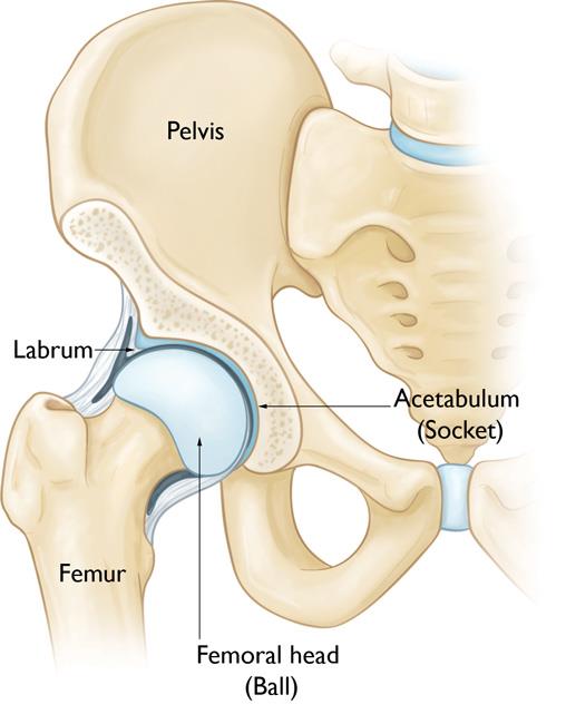
The hip is a ball-and-socket joint. The socket is formed by the acetabulum, which is part of the large pelvis bone. The ball is the femoral head, which is the upper end of the femur (thighbone).
A slippery tissue called articular cartilage covers the surface of the ball and the socket. It creates a smooth, low friction surface that helps the bones glide easily across each other during movement.
The acetabulum is ringed by strong fibrocartilage called the labrum. The labrum forms a gasket around the socket, creating a tight seal and helping to provide stability to the joint.
In FAI, bone overgrowth — called bone spurs — develop around the femoral head and/or along the acetabulum. This extra bone causes abnormal contact between the hip bones, and prevents them from moving smoothly during activity. Over time, this can result in tears of the labrum and the breakdown of articular cartilage (osteoarthritis).
There are three types of FAI: pincer, cam, and combined impingement.
FAI occurs because the hip bones do not form normally during the childhood growing years. It is the deformity of a cam bone spur, pincer bone spur, or both, that leads to joint damage and pain. When the hip bones are shaped abnormally, there is little that can be done to prevent FAI.
It is not known how many people have FAI. Some people may live long, active lives with FAI and never have problems. When symptoms develop, however, it usually indicates that there is damage to the cartilage or labrum and the disease is likely to progress.
Because athletic people may work the hip joint more vigorously, they may begin to experience pain earlier than those who are less active. However, exercise does not cause FAI.
The most common symptoms of FAI include:
Pain often occurs in the groin area, although it may occur toward the outside of the hip. Turning, twisting, and squatting may cause a sharp, stabbing pain. Sometimes, the pain is just a dull ache.
When symptoms first occur, it is helpful to try and identify an activity or something you may have done that could have caused the pain. Sometimes, you can modify your activities, let your hip rest, and see if the pain will settle down. Over-the-counter anti-inflammatory medicines, such as ibuprofen and naproxen, may help.
If your symptoms persist, you will need to see a doctor to determine the exact cause of your pain and provide treatment options. The longer painful symptoms go untreated, the more damage FAI can cause in the hip.
During your first appointment, your doctor will discuss your general health and your hip symptoms. He or she will also examine your hip.
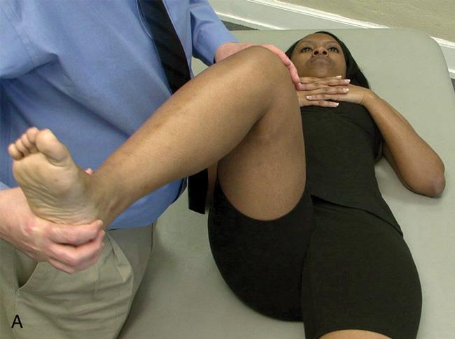
As part of the physical examination, your doctor will likely conduct the impingement test. For this test, your doctor will bring your knee up towards your chest and then rotate it inward towards your opposite shoulder. If this recreates your hip pain, the test result is positive for impingement.
Your doctor may order imaging tests to help determine whether you have FAI.
Activity changes. Your doctor may first recommend simply changing your daily routine and avoiding activities that cause symptoms.
Nonsteroidal anti-inflammatory medications. Drugs like ibuprofen can be provided in a prescription-strength form to help reduce pain and inflammation.
Physical therapy. Specific exercises can improve the range of motion in your hip and strengthen the muscles that support the joint. This can relieve some stress on the injured labrum or cartilage.
If tests show joint damage caused by FAI and your pain is not relieved by nonsurgical treatment, your doctor may recommend surgery.
Many FAI problems can be treated with arthroscopic surgery. Arthroscopic procedures are done with small incisions and thin instruments. The surgeon uses a small camera, called an arthroscope, to view inside the hip.
During arthroscopy, your doctor can repair or clean out any damage to the labrum and articular cartilage. He or she can correct the FAI by trimming the bony rim of the acetabulum and also shaving down the bump on the femoral head.
Some severe cases may require an open operation with a larger incision to accomplish this.
Surgery can successfully reduce symptoms caused by impingement. Correcting the impingement can prevent future damage to the hip joint. However, not all of the damage can be completely fixed by surgery, especially if treatment has been put off and the damage is severe. It is possible that more problems may develop in the future.
While there is a small chance that surgery might not help, it is currently the best way to treat painful FAI.
As the results of surgery improve, doctors will probably recommend earlier surgery for FAI. Surgical techniques continue to advance and in the future, computers may be used to help guide the surgeon in correcting and reshaping the hip.
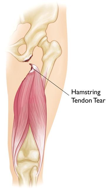
The hamstring muscles run down the back of the thigh. There are three hamstring muscles:
They start at the bottom of the pelvis at a place called the ischial tuberosity. They cross the knee joint and end at the lower leg. Hamstring muscle fibers join with the tough, connective tissue of the hamstring tendons near the points where the tendons attach to bones.
The hamstring muscle group helps you extend your leg straight back and bend your knee.
A hamstring strain can be a pull, a partial tear, or a complete tear.
Muscle strains are graded according to their severity. A grade 1 strain is mild and usually heals readily; a grade 3 strain is a complete tear of the muscle that may take months to heal.
Most hamstring injuries occur in the thick, central part of the muscle or where the muscle fibers join tendon fibers.
In the most severe hamstring injuries, the tendon tears completely away from the bone. It may even pull a piece of bone away with it. This is called an avulsion injury.
Muscle overload is the main cause of hamstring muscle strain. This can happen when the muscle is stretched beyond its capacity or challenged with a sudden load.
Hamstring muscle strains often occur when the muscle lengthens as it contracts, or shortens. Although it sounds contradictory, this happens when you extend a muscle while it is weighted, or loaded. This is called an “eccentric contraction.”
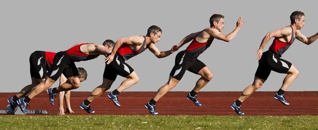
During sprinting, the hamstring muscles contract eccentrically as the back leg is straightened and the toes are used to push off and move forward. The hamstring muscles are not only lengthened at this point in the stride, but they are also loaded — with body weight as well as the force required for forward motion.
Like strains, hamstring tendon avulsions are also caused by large, sudden loads.
Several factors can make it more likely you will have a muscle strain, including:
Muscle tightness. Tight muscles are vulnerable to strain. Athletes should follow a year-round program of daily stretching exercises.
Muscle imbalance. When one muscle group is much stronger than its opposing muscle group, the imbalance can lead to a strain. This frequently happens with the hamstring muscles. The quadriceps muscles at the front of the thigh are usually more powerful. During high-speed activities, the hamstring may become fatigued faster than the quadriceps. This fatigue can lead to a strain.
Poor conditioning. If your muscles are weak, they are less able to cope with the stress of exercise and are more likely to be injured.
Muscle fatigue. Fatigue reduces the energy-absorbing capabilities of muscle, making them more susceptible to injury.
Choice of activity. Anyone can experience hamstring strain, but those especially at risk are:
Hamstring strains occur more often in adolescents because bones and muscles do not grow at the same rate. During a growth spurt, a child’s bones may grow faster than the muscles. The growing bone pulls the muscle tight. A sudden jump, stretch, or impact can tear the muscle away from its connection to the bone.
If you strain your hamstring while sprinting in full stride, you will notice a sudden, sharp pain in the back of your thigh. It will cause you to come to a quick stop, and either hop on your good leg or fall.
Additional symptoms may include:
People with hamstring strains often see a doctor because of a sudden pain in the back of the thigh that occurred when exercising.
During the physical examination, your doctor will ask about the injury and check your thigh for tenderness or bruising. He or she will palpate, or press, the back of your thigh to see if there is pain, weakness, swelling, or a more severe muscle injury.
Imaging tests that may help your doctor confirm your diagnosis include:
X-rays. An X-ray can show your doctor whether you have a hamstring tendon avulsion. This is when the injured tendon has pulled away a small piece of bone.
Magnetic Resonance Imaging (MRI). This study can create better images of soft tissues like the hamstring muscles. It can help your doctor determine the degree of your injury.
Treatment of hamstring strains will vary depending on the type of injury you have, its severity, and your own needs and expectations.
The goal of any treatment — nonsurgical or surgical — is to help you return to all the activities you enjoy. Following your doctor’s treatment plan will restore your abilities faster, and help you prevent further problems in the future.
Most hamstring strains heal very well with simple, nonsurgical treatment.
RICE. The RICE protocol is effective for most sports-related injuries. RICE stands for Rest, Ice, Compression, and Elevation.
Immobilization. Your doctor may recommend you wear a knee splint for a brief time. This will keep your leg in a neutral position to help it heal.
Physical therapy. Once the initial pain and swelling has settled down, physical therapy can begin. Specific exercises can restore range of motion and strength.
A therapy program focuses first on flexibility. Gentle stretches will improve your range of motion. As healing progresses, strengthening exercises will gradually be added to your program. Your doctor will discuss with you when it is safe to return to sports activity.
Surgery is most often performed for tendon avulsion injuries, where the tendon has pulled completely away from the bone. Tears from the pelvis (proximal tendon avulsions) are more common than tears from the shinbone (distal tendon avulsions).
Surgery may also be needed to repair a complete tear within the muscle.
Procedure. To repair a tendon avulsion, your surgeon must pull the hamstring muscle back into place and remove any scar tissue. Then the tendon is reattached to the bone using large stitches or staples.
A complete tear within the muscle is sewn back together using stitches.
Rehabilitation. After surgery, you will need to keep weight off of your leg to protect the repair. In addition to using crutches, you may need a brace that keeps your hamstring in a relaxed position. How long you will need these aids will depend on the type of injury you have.
Your physical therapy program will begin with gentle stretches to improve flexibility and range of motion. Strengthening exercises will gradually be added to your plan.
Rehabilitation for a proximal hamstring reattachment typically takes at least 6 months, due to the severity of the injury. Distal hamstring reattachments require approximately 3 months of rehabilitation before returning to athletic activities. Your doctor will tell you when it is safe to return to sports.
Most people who injure their hamstrings will recover full function after completing a rehabilitation plan. Early treatment with a plan that includes the RICE protocol and physical therapy has been shown to result in better function and quicker return to sports.
To prevent reinjuring your hamstring, be sure to follow your doctor’s treatment plan. Return to sports only after your doctor has given you the go-ahead. Reinjuring your hamstring increases your risk of permanent damage. This can result in a chronic condition.
Platelet-rich plasma (PRP) is currently being investigated for its effectiveness in speeding the healing of hamstring muscle injuries. PRP is a preparation developed from a patient’s own blood. It contains a high concentration of proteins called growth factors that are very important in the healing of injuries.
A few treatment centers across the country are incorporating PRP injections into the nonsurgical treatment regimen for some hamstring muscle injuries. However, this method is still under investigation and more research is necessary to prove PRP’s effectiveness.
The large bones that make up the hip joint—the femur (thighbone) and the pelvis—serve as anchors for several muscles. Some of these muscles move across the abdomen or the buttocks (hip flexors, gluteals). Others move down the thigh to the knee (abductors, adductors, quadriceps, hamstrings).
The strain may be a simple stretch in your muscle or tendon, or it may be a partial or complete tear of muscle fibers or of the muscle and tendon combination.
Once the muscle is injured, it becomes vulnerable to reinjury. Repeated strains in muscles about the hip and pelvis may be associated with athletic pubalgia (also called sports hernia). A sports hernia is a strain or tear of any soft tissue (muscle, tendon, ligament) in the lower abdomen or groin area. This condition is discussed in Sports Hernia (Athletic Pubalgia).
A hip strain can be an acute injury—meaning that it occurs suddenly, such as from a fall, a stretch injury, or a direct blow during contact sports. Hip strains are also caused by overuse—when the muscle or tendon has slowly become weakened over time by repetitive movements.
Factors that put you at greater risk for a hip strain include:
A muscle strain causes pain and tenderness in the injured area. Other symptoms may include:
Many hip strains will improve with simple home treatment. Mild strains can be treated with the RICE protocol. RICE stands for rest, ice, compression, and elevation.
In addition, nonsteroidal anti-inflammatory drugs, or NSAIDs, such as ibuprofen and naproxen, can help reduce swelling and relieve pain.
If the pain persists or it becomes more difficult to move your hip and leg, contact your doctor.
Your doctor will discuss your general health and ask you about what activities you were doing just prior to the injury. He or she will examine your leg and hip for tenderness or swelling. During the physical examination, your doctor will apply pressure to various muscles in the area and move your leg and hip in various directions to assess your range of motion.
Your doctor may also ask you to perform a variety of stretches and movements to help determine which muscle is injured.
X-rays provide images of dense structures such as bone. Your doctor may order an x-ray to rule out the possibility of a stress fracture of the hip, which has similar symptoms. In most cases, no additional imaging tests are needed to confirm the diagnosis.
Medical treatment for muscle strains is designed to relieve pain and restore range of motion and strength. The majority of hip strains are treated nonsurgically.
In addition to the RICE method and anti-inflammatory medication, your doctor may recommend using crutches for a few days to limit the weight on your hip. Other recommendations may include:
Severe injuries in which the tendons are completely avulsed from bone may require surgery in order to return to normal function and movement. Surgery typically involves re-attaching the torn tendon tissue back to the bone.
It is important to know that many severe hip strains are successfully treated without surgery. Your doctor will discuss the treatment options that best meet your individual health needs.
In most cases, you should avoid the activity that caused your injury for 10 to14 days. A severe muscle strain may require a longer period of recovery. If your pain returns when you resume more strenuous activity, however, discontinue what you are doing and go back to easier activities that do not cause pain.
You can take the following precautions to help prevent muscle strains in the future:
Contusions occur when a direct blow or repeated blows by a blunt object strike part of the body, crushing underlying muscle fibers and connective tissue without breaking the skin. A contusion can result from falling or jamming the body against a hard surface.
Contusions cause swelling and pain and can limit joint range of motion near the injury. Torn blood vessels may cause bluish discoloration. The injured muscle may feel weak and stiff.
Sometimes a pool of blood collects within damaged tissue, forming a lump over the injury (hematoma). If tissue damage is extensive, you may also have a broken bone, dislocated joint, sprain, torn muscle, or other injuries.
Contusions to the abdomen may damage internal organs.
See your doctor right away for complete diagnosis. A physical examination will determine the exact location and extent of the injury.
Diagnostic imaging tools may be used to better visualize inside the injured area of your body. These tools include x-rays, ultrasound, magnetic resonance imaging (MRI) scans, or computerized tomography (CT) scans.
For some injuries, your doctor may also need to check for nerve injury.
To control pain, bleeding, and inflammation, keep the muscle in a gentle stretch position and use the RICE protocol:
Most athletes with contusions get better quickly with simple treatment measures. Your doctor may give you nonsteroidal anti-inflammatory drugs (NSAIDs), such as ibuprofen, or other medications for pain relief. Do not massage the injured area.
During the first 24 to 48 hours after injury (acute phase), you will probably need to continue using rest, ice, compression bandages, and elevation of the injured area to control bleeding, swelling, and pain. While the injured muscle heals, be sure to keep exercising the uninjured parts of your body to maintain your overall level of fitness.
If there is a large hematoma that does not go away within several days, your doctor may drain it surgically to expedite healing.
After a few days, inflammation and swelling should start to decrease and the injury may feel a little better. At this time, your doctor may tell you to apply gentle heat to the injury and start the rehabilitation process. Remember to increase your activity level gradually.
Depending upon the extent of your injuries, returning to your normal sports activity may take several weeks or longer. If you put too much stress on the injured area before it has healed enough, excessive scar tissue may develop and cause more problems.
In the first phase of rehabilitation, your doctor may prescribe gentle stretching exercises that begin to restore range of motion to the injured area.
Once your range of motion has improved, your doctor will likely recommend incorporating weightbearing and strengthening exercises.
When you have normal, pain-free range of motion, your doctor may let you return to non-contact sports.
You may be able to return to contact sports when you get back your full strength, motion, and endurance. When your doctor and athletic trainer agree that you are ready to return to play, they may want you to wear a customized protective device to prevent further injury to the area that had a contusion.
Depending upon your sport, you may get special padding made of firm or semi-firm materials. The padding spreads out the force of impact when direct blows from blunt objects strike your body.
Getting prompt medical treatment and following your doctor’s advice about rehabilitation can help you avoid serious medical complications that occasionally result from deep muscle contusions. Two of the more common complications are compartment syndrome and myositis ossificans.
In certain cases, rapid bleeding may cause extremely painful swelling within the muscles of your arm, leg, foot, or buttock. Build-up of pressure from fluids several hours after a contusion can disrupt blood flow and prevent nourishment from reaching the muscle group. Compartment syndrome may require urgent surgery to relieve the pressure on the muscles, blood vessels, and nerves.
Young athletes who try to rehabilitate a severe contusion too quickly sometimes develop myositis ossificans. This is a condition in which bone forms in the injured muscle.
Symptoms may include mild to severe pain that does not go away and swelling at the site of the injury. Abnormal bone formation can also reduce your flexibility. Vigorous stretching exercises may make the condition worse.
Rest, ice, compression, and elevation to reduce inflammation will usually help. Gentle stretching exercises may improve flexibility. Surgery is rarely required.
The most commonly affected muscle groups are:
Cramps in the feet, hands, arms, abdomen, and along the rib cage are also very common.
Although the exact cause of muscle cramps is unknown (idiopathic), some researchers believe inadequate stretching and muscle fatigue leads to abnormalities in the mechanisms that control muscle contraction. Other factors may also be involved, including poor conditioning, exercising or working in intense heat, dehydration and depletion of salt and minerals (electrolytes).
Muscles are bundles of fibers that contract and expand to produce movement. A regular program of stretching lengthens muscle fibers so they can contract and tighten more vigorously when you exercise. When your body is poorly conditioned, you are more likely to experience muscle fatigue, which can alter spinal neural reflex activity. Overexertion depletes a muscle’s oxygen supply, leading to build up of waste product and spasm. When a cramp begins, the spinal cord stimulates the muscle to keep contracting.
Muscle cramps are more likely when you exercise in hot weather because sweat drains your body’s fluids, salt and minerals (i.e., potassium, magnesium and calcium). Loss of these nutrients may also cause a muscle to spasm.
Some people are predisposed to muscle cramps and get them regularly with any physical exertion.
Those at greatest risk for cramps and other ailments related to excess heat include infants and young children, and people over age 65. Other factors that put people at greater risk for muscle cramp include:
Muscle cramps are very common among endurance athletes, such as marathon runners and triathletes, and older people who perform strenuous physical activities.
Muscle cramps range in intensity from a slight tic to agonizing pain. A cramping muscle may feel hard to the touch and/or appear visibly distorted or twitch beneath the skin. A cramp can last a few seconds to 15 minutes or longer. It might recur multiple times before it goes away.
Cramps usually go away on their own without seeing a doctor.
To avoid future cramps, work toward better overall fitness. Do regular flexibility exercises before and after you work out to stretch muscle groups most prone to cramping.
Always warm up before stretching. Good examples of warm-up activities are slowly running in place or walking briskly for a few minutes.
Lean forward against a wall with one leg in front of the other. Straighten your back leg and press your heel into the floor. Your front knee is bent. Hold for 15 to 30 seconds.
Do: Keep both heels flat on the floor. Point the toes of your back foot toward the heel of your front foot.
Sit up tall with both legs extended straight in front of you. Your feet are neutral — not pointed or flexed. Place your palms on the floor and slide your hands toward your ankles. Hold for 30 seconds.
Do: Keep your chest open and back long. Reach from your hips. Stop sliding your palms forward when you feel the stretch.
Do not: Round your back or try to bring your nose to your knees. Do not lock your knees.
Hold on to a wall or the back of a chair for balance. Lift one foot and bring your heel up toward your buttocks. Grasp your ankle with your hand and pull your heel closer to your body. Hold the stretch for 30 seconds.
Do: Keep your knees close together. Stop bringing your heel closer when you feel the stretch.
Do not: Arch or twist your back.
Hold each stretch briefly, then release. Never stretch to the point of pain.
Although most muscle cramps are benign, sometimes they can indicate a serious medical condition.
See your doctor if cramps are severe, happen frequently, respond poorly to simple treatments, or are not related to obvious causes like strenuous exercise. You could have problems with circulation, nerves, metabolism, hormones, medications, or nutrition.
Muscle cramps may be a part of many conditions that range from minor to severe, such as Lou Gehrig’s disease (amyotrophic lateral sclerosis), spinal nerve irritation or compression (radiculopathy), hardening of the arteries, narrowing of the spinal canal (stenosis), thyroid disease, chronic infections, and cirrhosis of the liver.
Overuse injuries occur in a wide range of sports, from baseball and basketball to track, soccer, and gymnastics. Some of these injuries are unique to a certain sport, such as throwing injuries of the elbow and shoulder that are prevalent in baseball players. The most common overuse injuries involve the knee and foot.
Overuse injuries can affect muscles, ligaments, tendons, bones, and growth plates. In children, these structures are still growing, and the growth is generally uneven. Bones grow faster than muscles in children. This uneven growth pattern makes younger athletes more susceptible to muscle, tendon, and growth plate injuries.
Growth plates are the areas of developing cartilage where bone growth occurs in children. The growth plates are weaker than the nearby ligaments and tendons. Repetitive stress can lead to injury of the growth plate and disrupt the normal growth of the bone.
As organized youth athletics has grown in popularity, the pressure to compete has led to children specializing in one sport only. In generations past, children changed sports with the seasons throughout the year, but today it is common for a child to play just one sport year-round. Many children play on more than one team at the same time, as well.
When a child participates in just one sport throughout the year, he or she continually uses the same muscle groups and applies unchanging stress to specific areas of the body. This can lead to muscle imbalances that, when combined with overtraining and inadequate periods of rest, put children at serious risk for overuse injuries.
Coaches and parents should be aware of the more common signs of overuse injury. These include:
Sever’s disease is one of most common causes of heel pain in children, and often occurs during adolescence when children hit a growth spurt. Also known as an osteochondrosis or apophysitis, Sever’s disease is an inflammatory condition of the growth plate in the heel bone (calcaneus). Running, jumping, and being active lead to repetitive stress on the growth plate as the foot strikes the ground. This results in inflammation (swelling) in the growth plate which causes heel pain.
The primary treatment of Sever’s disease is to rest the foot (stop the sport) until the pain goes away. Once the pain is gone, the child may return to normal activities.
Additional treatments may include:
In cases where the pain is bad enough to interfere with walking, a short-leg cast or “walker boot” might be required to immobilize the foot while it heals.
It is not unusual for Sever’s disease to recur. This typically happens when a child increases sports activities. Wearing sports shoes that provide good support to the foot and heel may help prevent recurrence. Sever’s disease will not return once a child is fully grown and the growth plate in the heel has hardened into bone.
In Osgood-Schlatter disease, children have pain at the front of the knee due to inflammation of the growth plate at the upper end of the tibia (shinbone). This growth plate (known as the tibial tubercle) is a bump near the top of the tibia where the tendon from the kneecap (patellar tendon) attaches to the bone.
When a child is active, the quadriceps muscles of the thigh pull on the patellar tendon which in turn, pulls on the tibial tubercle. In some children, this repetitive traction on the tubercle leads to inflammation, swelling, and tenderness. The prominence, or bump, of the tibial tubercle may become very pronounced. Painful symptoms are often brought on by running, jumping, and other sports-related activities.
Treatment of Osgood-Schlatter disease focuses on limiting exercise activity until the pain resolves.
Most symptoms will completely disappear when a child completes the adolescent growth spurt, around age 14 for girls and age 16 for boys. However, the prominence of the tubercle will persist.
Until a child is fully grown, however, Osgood-Schlatter disease can lead to more severe problems if it is not allowed to heal. Continued stress on the tibial tubercle from athletic activity could potentially lead to a break in the tubercle bone. Treatment for this type of fracture will require casting or even surgery. In order for the fracture to safely heal, a child will not be allowed to participate in sports for a prolonged period of time.
Jumper’s Knee
Although these types of problems in the kneecap commonly occur in children who are active in sports that involve jumping — such as basketball and volleyball — they can be seen in almost any sport.
Whether the problem stems from the tendon or growth plate, a child will need to rest from sports activities until the pain resolves.
In addition to rest, your doctor may recommend applying an ice pack to the knee every 2 to 3 hours for a few days until the pain starts to go away. Nonsteroidal anti-inflammatory medications may also help to relieve painful symptoms.
Like Osgood-Schlatter disease, jumper’s knee can result in a fracture if a child continues to participate in sports activity without adequate rest. A fracture at the lower end of the kneecap will require treatment with casting or surgery, depending upon the type of fracture. The recovery time for a fracture of the patella requires prolonged absence from sports.
A child’s elbow can be injured from repetitive overhand throwing, as seen with pitching in baseball. Although throwing injuries in the elbow most commonly occur in pitchers, they can be seen in any child who participates in repetitive overhand throwing.
The overhand throw creates stresses on the growth areas of the immature elbow. If overdone, overhand throwing may result in excessive inflammation of structures in the elbow, such as ligaments, cartilage, and growth plates.
Medial apophysitis. One of the most common elbow problems in active children is medial apophysitis, often referred to as “Little Leaguer’s elbow.” Medial apophysitis causes pain at the bony bump on the inside of the elbow. The bump, called the medial epicondyle, is at the end of the humerus bone and contains a growth plate called the medial apophysis. Muscles that control wrist motion attach to the medial epicondyle, and excessive overhand throwing can irritate and inflame the growth plate.
Osteochondritis dissecans. A common source of pain at the outside of the elbow in active children is osteochondritis dissecans. Excessive overhand throwing can compress the immature bones of the elbow joint, causing small pieces of cartilage and bone to loosen and float inside the joint.
As with all overuse injuries, the key to pain relief is resting from the sport. If pain continues after a few days of complete rest, seek medical treatment. If left untreated, throwing injuries in the elbow can become complicated conditions. Depending upon the severity of a child’s injury, surgery may be required.
Stress fractures occur when muscles become fatigued and transfer the overload of stress to bones. Bone is a living tissue and is constantly in a balanced cycle of building up and breaking down. If a child overdoes activity and too much stress is being placed on bones, the body will not be able to build up bone fast enough. Eventually, the bone fails and a small crack (stress fracture) develops.
The repetitive force that causes a stress fracture is not great enough to cause an acute fracture — such as a broken arm caused by a fall. Most stress fractures occur in the weightbearing bones of the lower leg and the foot. They often result from a sudden increase in activity, such as when a child begins cross country running after a summer of rest.
Stress fractures typically require 6 to 8 weeks to heal. During that time, the child cannot participate in the activity that caused the injury. A cast or brace may be needed to rest the injured bones while they heal. In some cases, a rehabilitation protocol to return to sport may also be recommended.
If the activity that caused the stress fracture is resumed too quickly, larger, harder-to-heal stress fractures can develop. Re-injury also could lead to chronic problems where the stress fracture might never heal properly.
Repetitive stress on a growth plate (physis) in the arms or legs can be painful and, if ignored, can impair growth.
Overuse stress reaction leads to irregularity or widening of the growth plate. If repetitive stress continues, the growth plate can become permanently damaged and may actually stop growing prematurely. This could lead to a deformity.
Common examples of sports activities that may cause stress reaction in growth plates include gymnastics and overhand throwing. Gymnasts perform repetitive wrist activities that can lead to stress reaction of the growth plate in the distal radius bone. Young baseball pitchers apply forces across the upper arm bone in their shoulders during the overhand throw. The growth plate in the upper humerus of the shoulder can be damaged and appear widened in an x-ray image.
If the growth plate appears abnormal in an x-ray of a child who participates in a high-risk sport, treatment requires stopping the aggravating activity for 2 to 3 months.
Soft tissues such as muscles, ligaments, and tendons can be injured from overuse. Just about every sports activity can be associated with this.
A period of rest from the sport and nonsteroidal anti-inflammatory medicine will typically relieve pain associated with strains and sprains. Physical therapy exercises may be recommended to help restore strength and mobility.
Many overuse injuries in children can be prevented. Key to prevention is to avoid overdoing any single sport, and to give growing bodies adequate rest between practices or games.
The American Academy of Orthopaedic Surgeons has partnered with STOP Sports Injuries to help educate parents, coaches, and athletes about how to prevent overuse injuries. Specific tips to prevent overuse injuries include:
Shin splints (medial tibial stress syndrome) is an inflammation of the muscles, tendons, and bone tissue around your tibia. Pain typically occurs along the inner border of the tibia, where muscles attach to the bone.
In general, shin splints develop when the muscle and bone tissue (periosteum) in the leg become overworked by repetitive activity.
Shin splints often occur after sudden changes in physical activity. These can be changes in frequency, such as increasing the number of days you exercise each week. Changes in duration and intensity, such as running longer distances or on hills, can also cause shin splints.
Other factors that contribute to shin splints include:
Runners are at highest risk for developing shin splints. Dancers and military recruits are two other groups frequently diagnosed with the condition.
The most common symptom of shin splints is pain along the border of the tibia. Mild swelling in the area may also occur.
Shin splint pain may:
After discussing your symptoms and medical history, your doctor will examine your lower leg. An accurate diagnosis is very important. Sometimes, other problems may exist that can have an impact on healing.
Your doctor may order additional imaging tests to rule out other shin problems. Several conditions can cause shin pain, including stress fractures, tendinitis, and chronic exertional compartment syndrome.
If your shin splints are not responsive to treatment, your doctor may want to make sure you do not have a stress fracture. A stress fracture is a small crack(s) in the tibia caused by stress and overuse.
Imaging tests that create pictures of anatomy help to diagnose conditions. A bone scan and magnetic resonance imaging (MRI) study will often show stress fractures in the tibia.
Tendons attach muscles to bones. Tendinitis occurs when tendons become inflamed. This can be painful like shin splints, especially if there is a partial tear of the involved tendon. An MRI can help diagnose tendinitis.
An uncommon condition called chronic exertional compartment syndrome causes symptoms like shin splints. Compartment syndrome is a painful condition that occurs when pressure within the muscles builds to dangerous levels. In chronic exertional compartment syndrome, this is brought on by exercise. Pain usually resolves soon after the activity stops.
The tests used to diagnose this condition involve measuring the pressure within the leg compartments before and after exercise.
Rest. Because shin splints are typically caused by overuse, standard treatment includes several weeks of rest from the activity that caused the pain. Lower impact types of aerobic activity can be substituted during your recovery, such as swimming, using a stationary bike, or an elliptical trainer.
Nonsteroidal anti-inflammatory medicines. Drugs like ibuprofen, aspirin, and naproxen reduce pain and swelling.
Ice. Use cold packs for 20 minutes at a time, several times a day. Do not apply ice directly to the skin.
Compression. Wearing an elastic compression bandage may prevent additional swelling.
Flexibility exercises. Stretching your lower leg muscles may make your shins feel better.
Supportive shoes. Wearing shoes with good cushioning during daily activities will help reduce stress in your shins.
Orthotics. People who have flat feet or recurrent problems with shin splints may benefit from orthotics. Shoe inserts can help align and stabilize your foot and ankle, taking stress off of your lower leg. Orthotics can be custom-made for your foot, or purchased “off the shelf.”
Return to exercise. Shin splints usually resolve with rest and the simple treatments described above. Before returning to exercise, you should be pain-free for at least 2 weeks. Keep in mind that, when you return to exercise, it must be at a lower level of intensity. You should not be exercising as often as you did before, or for the same length of time.
Be sure to warm up and stretch thoroughly before you exercise. Increase training slowly. If you start to feel the same pain, stop exercising immediately. Use a cold pack and rest for a day or two. Return to training again at a lower level of intensity. Increase training even more slowly than before.
Very few people need surgery for shin splints. Surgery has been done in very severe cases that do not respond to nonsurgical treatment. It is not clear how effective surgery is, however.
There are things you can do to prevent shin splints.
Wear a proper fitting athletic shoe. To get the right fit, determine the shape of your foot using the “wet test.” Step out of the shower onto a surface that will show your footprint, like a brown paper bag. If you have a flat foot, you will see an impression of your whole foot on the paper. If you have a high arch, you will only see the ball and heel of your foot. When shopping, look for athletic shoes that match your particular foot pattern.
In addition, make sure you wear shoes designed for your sport. Running long distances in court-type sneakers can contribute to shin splints.
Slowly build your fitness level. Increase the duration, intensity, and frequency of your exercise regimen gradually.
Cross train. Alternate jogging with lower impact sports like swimming or cycling.
Barefoot running. In recent years, barefoot running has gained in popularity. Many people claim it has helped to resolve shin splints. Some research indicates that barefoot running spreads out impact stresses among muscles, so that no area is overloaded. However, there is no clear evidence that barefoot running reduces the risk for any injury.
Like any significant change in your fitness regimen, a barefoot running program should be started very gradually. Begin with short distances to give your muscles and your feet time to adjust. Pushing too far, too fast can put you at risk for stress injuries. In addition, barefoot runners are at increased risk for cuts and bruises on their feet. Several brands of minimalist shoes with “toes” are available and these also require a slow working in period as your body adjusts to this different activity.
If your shin splints do not improve after rest and other methods described above, be sure to see a doctor to determine whether something else is causing your leg pain.
The plantar fascia is a long, thin ligament that lies directly beneath the skin on the bottom of your foot. It connects the heel to the front of your foot, and supports the arch of your foot.
The plantar fascia is designed to absorb the high stresses and strains we place on our feet. But, sometimes, too much pressure damages or tears the tissues. The body’s natural response to injury is inflammation, which results in the heel pain and stiffness of plantar fasciitis.
In most cases, plantar fasciitis develops without a specific, identifiable reason. There are, however, many factors that can make you more prone to the condition:
Although many people with plantar fasciitis have heel spurs, spurs are not the cause of plantar fasciitis pain. One out of 10 people has heel spurs, but only 1 out of 20 people (5%) with heel spurs has foot pain. Because the spur is not the cause of plantar fasciitis, the pain can be treated without removing the spur.
Related Articles
Warm Up, Cool Down and Be Flexible
Orthotics
Athletic Shoes
Stress Fractures of the Foot and Ankle
The most common symptoms of plantar fasciitis include:
After you describe your symptoms and discuss your concerns, your doctor will examine your foot. Your doctor will look for these signs:
Your doctor may order imaging tests to help make sure your heel pain is caused by plantar fasciitis and not another problem.
X-rays provide clear images of bones. They are useful in ruling out other causes of heel pain, such as fractures or arthritis. Heel spurs can be seen on an x-ray.
Other imaging tests, such as magnetic resonance imaging (MRI) and ultrasound, are not routinely used to diagnose plantar fasciitis. They are rarely ordered. An MRI scan may be used if the heel pain is not relieved by initial treatment methods.
More than 90% of patients with plantar fasciitis will improve within 10 months of starting simple treatment methods.
Rest. Decreasing or even stopping the activities that make the pain worse is the first step in reducing the pain. You may need to stop athletic activities where your feet pound on hard surfaces (for example, running or step aerobics).
Ice. Rolling your foot over a cold water bottle or ice for 20 minutes is effective. This can be done 3 to 4 times a day.
Nonsteroidal anti-inflammatory medication. Drugs such as ibuprofen or naproxen reduce pain and inflammation. Using the medication for more than 1 month should be reviewed with your primary care doctor.
Exercise. Plantar fasciitis is aggravated by tight muscles in your feet and calves. Stretching your calves and plantar fascia is the most effective way to relieve the pain that comes with this condition.
Cortisone injections. Cortisone, a type of steroid, is a powerful anti-inflammatory medication. It can be injected into the plantar fascia to reduce inflammation and pain. Your doctor may limit your injections. Multiple steroid injections can cause the plantar fascia to rupture (tear), which can lead to a flat foot and chronic pain.
Supportive shoes and orthotics. Shoes with thick soles and extra cushioning can reduce pain with standing and walking. As you step and your heel strikes the ground, a significant amount of tension is placed on the fascia, which causes microtrauma (tiny tears in the tissue). A cushioned shoe or insert reduces this tension and the microtrauma that occurs with every step. Soft silicone heel pads are inexpensive and work by elevating and cushioning your heel. Pre-made or custom orthotics (shoe inserts) are also helpful.
Night splints. Most people sleep with their feet pointed down. This relaxes the plantar fascia and is one of the reasons for morning heel pain. A night splint stretches the plantar fascia while you sleep. Although it can be difficult to sleep with, a night splint is very effective and does not have to be used once the pain is gone.
Physical therapy. Your doctor may suggest that you work with a physical therapist on an exercise program that focuses on stretching your calf muscles and plantar fascia. In addition to exercises like the ones mentioned above, a physical therapy program may involve specialized ice treatments, massage, and medication to decrease inflammation around the plantar fascia.
Extracorporeal shockwave therapy (ESWT). During this procedure, high-energy shockwave impulses stimulate the healing process in damaged plantar fascia tissue. ESWT has not shown consistent results and, therefore, is not commonly performed.
ESWT is noninvasive—it does not require a surgical incision. Because of the minimal risk involved, ESWT is sometimes tried before surgery is considered.
Surgery is considered only after 12 months of aggressive nonsurgical treatment.
Gastrocnemius recession. This is a surgical lengthening of the calf (gastrocnemius) muscles. Because tight calf muscles place increased stress on the plantar fascia, this procedure is useful for patients who still have difficulty flexing their feet, despite a year of calf stretches.
In gastrocnemius recession, one of the two muscles that make up the calf is lengthened to increase the motion of the ankle. The procedure can be performed with a traditional, open incision or with a smaller incision and an endoscope, an instrument that contains a small camera. Your doctor will discuss the procedure that best meets your needs.
Complication rates for gastrocnemius recession are low, but can include nerve damage.
Plantar fascia release. If you have a normal range of ankle motion and continued heel pain, your doctor may recommend a partial release procedure. During surgery, the plantar fascia ligament is partially cut to relieve tension in the tissue. If you have a large bone spur, it will be removed, as well. Although the surgery can be performed endoscopically, it is more difficult than with an open incision. In addition, endoscopy has a higher risk of nerve damage.
Complications. The most common complications of release surgery include incomplete relief of pain and nerve damage.
Recovery. Most patients have good results from surgery. However, because surgery can result in chronic pain and dissatisfaction, it is recommended only after all nonsurgical measures have been exhausted.
The hip is a ball-and-socket joint formed where the rounded end of the thighbone (femur) fits into a cup-shaped socket (acetabulum) in the pelvis. The acetabulum is ringed by strong fibrocartilage called the labrum that creates a tight seal and helps to provide stability to the joint.
Encasing the hip are ligaments that surround the joint and hold it together. Over the ligaments are tendons that attach muscles in the buttocks, thighs, and pelvis to the bones. These muscles control hip movement.
Fluid-filled sacs called bursae are located in strategic spots around the hip to provide cushioning and help the muscles move smoothly over the bone.
Snapping hip can occur in different areas of the hip where tendons and muscles slide over knobs in the hip bones.
Snapping hip is most often the result of tightness in the muscles and tendons surrounding the hip. People who are involved in sports and activities that require repeated bending at the hip are more likely to experience snapping hip. Dancers are especially vulnerable.
Young athletes are also more likely to have snapping hip. This is because tightness in the muscle structures of the hip is common during adolescent growth spurts.
Most people do not see a doctor for snapping hip unless they experience some pain. If the snapping hip bothers you — but not to the point of seeing a doctor — try the following conservative home treatment options:
If you are still experiencing discomfort after trying these conservative methods, consult your doctor for professional treatment.
Your doctor will first determine the exact cause of the snapping by discussing your medical history and symptoms, and conducting a physical examination. He or she may ask you where it hurts, what kinds of activities bring on the snapping, whether you can demonstrate the snapping, or whether you have experienced any injury to the hip area.
You may also be asked to stand and move your hip in various directions to reproduce the snapping. Your doctor may even be able to feel the tendon moving as you bend or extend your hip.
X-rays provide clear pictures of dense structures, such as bone. Although x-rays of people with snapping hip do not typically show anything abnormal, your doctor may order x-rays or other tests to rule out any problems with the bones or joint.
Initial treatment typically involves a period of rest and modification of activities. Depending upon the cause of your snapping hip, your doctor may also recommend other conservative treatment options.
Your doctor may prescribe exercises like the ones below to stretch and strengthen the musculature surrounding the hip. Guidance from a physical therapist may also be recommended.
If you have hip bursitis, your doctor may recommend an injection of a corticosteroid into the bursa to reduce painful inflammation.
In the rare instances that snapping hip does not respond to conservative treatment, your doctor may recommend surgery. The type of surgery will depend on the cause of the snapping hip.
Your orthopaedic surgeon will discuss with you the best procedure to meet your individual health needs.
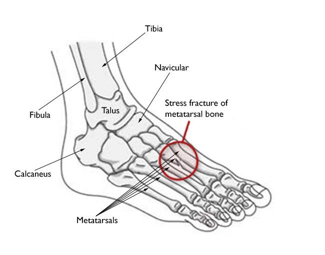
Stress fractures occur most often in the second and third metatarsals in the foot, which are thinner (and often longer) than the adjacent first metatarsal. This is the area of greatest impact on your foot as you push off when you walk or run.
Stress fractures are also common in the calcaneus (heel); fibula (the outer bone of the lower leg and ankle); talus (a small bone in the ankle joint); and the navicular (a bone on the top of the midfoot).
Many stress fractures are overuse injuries. They occur over time when repetitive forces result in microscopic damage to the bone. The repetitive force that causes a stress fracture is not great enough to cause an acute fracture — such as a broken ankle caused by a fall. Overuse stress fractures occur when an athletic movement is repeated so often, weight-bearing bones and supporting muscles do not have enough time to heal between exercise sessions.
Bone is in a constant state of turnover—a process called remodeling. New bone develops and replaces older bone. If an athlete’s activity is too great, the breakdown of older bone occurs rapidly — it outpaces the body’s ability to repair and replace it. As a result, the bone weakens and becomes vulnerable to stress fractures.
The most common cause of stress fractures is a sudden increase in physical activity. This increase can be in the frequency of activity—such as exercising more days per week. It can also be in the duration or intensity of activity—such as running longer distances.
Even for the nonathlete, a sudden increase in activity can cause a stress fracture. For example, if you walk infrequently on a day-to-day basis but end up walking excessively (or on uneven surfaces) while on a vacation, you might experience a stress fracture. A new style of shoes can lessen your foot’s ability to absorb repetitive forces and result in a stress fracture.
Conditions that decrease bone strength and density, such as osteoporosis, and certain long-term medications can make you more likely to experience a stress fracture-even when you are performing normal everyday activities. For example, stress fractures are more common in the winter months, when Vitamin D is lower in the body.
Studies show that female athletes are more prone to stress fractures than male athletes. This may be due, in part, to decreased bone density from a condition that doctors call the “female athlete triad.” When a girl or young woman goes to extremes in dieting or exercise, three interrelated illnesses may develop: eating disorders, menstrual dysfunction, and premature osteoporosis. As a female athlete’s bone mass decreases, her chances for getting a stress fracture increase.
Doing too much too soon is a common cause of stress fracture. This is often the case with individuals who are just beginning an exercise program-but it occurs in experienced athletes, as well. For example, runners who train less over the winter months may be anxious to pick up right where they left off at the end of the previous season. Instead of starting off slowly, they resume running at their previous mileage. This situation in which athletes not only increase activity levels, but push through any discomfort and do not give their bodies the opportunity to recover, can lead to stress fractures.
Anything that alters the mechanics of how your foot absorbs impact as it strikes the ground may increase your risk for a stress fracture. For example, if you have a blister, bunion, or tendonitis, it can affect how you put weight on your foot when you walk or run, and may require an area of bone to handle more weight and pressure than usual.
A change in training or playing surface, such as a tennis player going from a grass court to a hard court, or a runner moving from a treadmill to an outdoor track, can increase the risk for stress fracture.
Wearing worn or flimsy shoes that have lost their shock-absorbing ability may contribute to stress fractures.
The most common symptom of a stress fracture in the foot or ankle is pain. The pain usually develops gradually and worsens during weight-bearing activity. Other symptoms may include:
See your doctor as soon as possible if you think that you have a stress fracture in your foot or ankle. Ignoring the pain can have serious consequences. The bone may break completely.
Until your appointment with the doctor, follow the RICE protocol. RICE stands for rest, ice, compression, and elevation.
In addition, nonsteroidal anti-inflammatory drugs, or NSAIDs, such as ibuprofen or naproxen can help relieve pain and reduce swelling.
Your doctor will discuss your medical history and general health. He or she will ask about your work, your activities, your diet, and what medications you are taking.It is important that your doctor is aware of your risk factors for stress fracture. If you have had a stress fracture before, your doctor may order a full medical work-up with laboratory tests to check for nutritional deficiencies such as low calcium or Vitamin D.
After discussing your symptoms and health history, your doctor will examine your foot and ankle. During the examination, he or she will look for areas of tenderness and apply gentle pressure directly to the injured bone. Often, the key to diagnosing a stress fracture is the patient’s report of pain in response to this pressure. Pain from a stress fracture is typically limited to the area directly over the injured bone and is not generalized over the whole foot.
Your doctor may order imaging tests to help confirm the diagnosis.
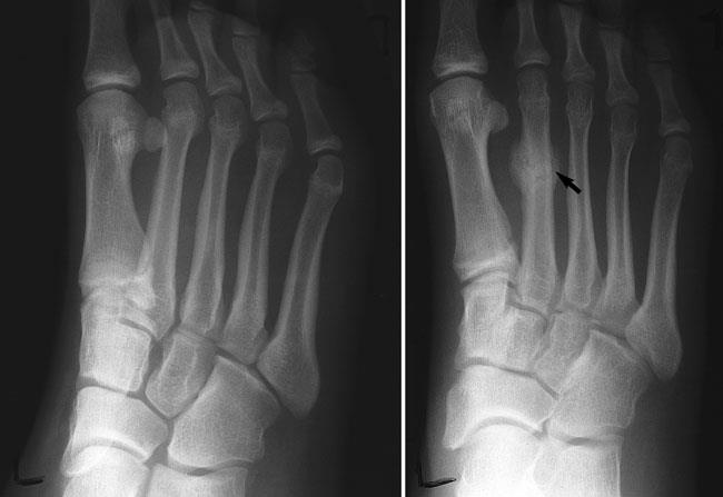
X-rays. X-rays provide images of dense structures, such as bone. Since a stress fracture starts as a tiny crack, it is often difficult to see on a first x-ray. The fracture may not be visible until several weeks later when it has actually started to heal. After a few weeks, a type of healing bone called callus may appear around the fracture site. In many cases, this is the point at which the fracture line actually becomes visible in the bone.
Other imaging studies. If your doctor suspects a stress fracture but cannot see it on an x-ray, he or she may recommend a bone scan or a magnetic resonance imaging (MRI) scan. Although a bone scan is less specific than an MRI in showing the actual location of the stress fracture, both of these types of studies are more sensitive than x-rays and can detect stress fractures earlier.
The goal of treatment is to relieve pain and allow the fracture to heal so that you are able to return to your activities. Following your doctor’s treatment plan will help you return to activities faster and prevent further damage to the bone.
Treatment will vary depending on the location of the stress fracture and its severity. The majority of stress fractures are treated nonsurgically.
In addition to the RICE protocol and anti-inflammatory medication, your doctor may recommend that you use crutches to keep weight off your foot until the pain subsides. Other recommendations for nonsurgical treatment may include:
Modified activities. It typically takes from 6 to 8 weeks for a stress fracture to heal. During that time, switch to activities that place less stress on your foot and leg. Swimming and cycling are good alternative activities. However, you should not resume any type of physical activity that involves your injured foot or ankle-even if it is low impact-without your doctor’s recommendation.
Protective footwear. To reduce stress on your foot and leg, your doctor may recommend wearing protective footwear. This may be a stiff-soled shoe, a wooden-soled sandal, or a removable short-leg fracture brace shoe.
Casting. Stress fractures in the fifth metatarsal bone (on the outer side of the foot) or in the navicular or talus bones take longer to heal. Your doctor may apply a cast to your foot to keep your bones in a fixed position and to remove the stress on your involved leg.
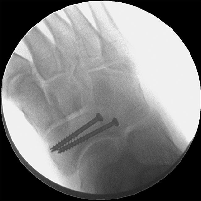
Some stress fractures require surgery to heal properly. In most cases, this involves supporting the bones by inserting a type of fastener. This is called internal fixation. Pins, screws, and/or plates are most often used to hold the small bones of the foot and ankle together during the healing process.
In most cases, it takes from 6 to 8 weeks for a stress fracture to heal. More serious stress fractures can take longer. Although it can be hard to be sidelined with an injury, returning to activity too soon can put you at risk for larger, harder-to-heal stress fractures and an even longer down time. Reinjury could lead to chronic problems and the stress fracture might never heal properly.
Once your pain has subsided, your doctor may confirm that the stress fracture has healed by taking x-rays. A computed tomography (CT) scan can also be useful in determining healing, especially in bones where the fracture line was initially hard to see.
Once the stress fracture has healed and you are pain free, your doctor will allow a gradual return to activity. During the early phase of rehabilitation, your doctor may recommend alternating days of activity with days of rest. This gives your bone the time to grow and withstand the new demands being placed upon it. As your fitness level improves, slowly increase the frequency, duration, and intensity of your exercise.
The following guidelines can help you prevent stress fractures in the future:
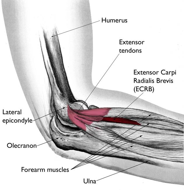
Your elbow joint is a joint made up of three bones: your upper arm bone (humerus) and the two bones in your forearm (radius and ulna). There are bony bumps at the bottom of the humerus called epicondyles, where several muscles of the forearm begin their course. The bony bump on the outside (lateral side) of the elbow is called the lateral epicondyle.
Muscles, ligaments, and tendons hold the elbow joint together.
Lateral epicondylitis, or tennis elbow, involves the muscles and tendons of your forearm that are responsible for the extension of your wrist and fingers. Your forearm muscles extend your wrist and fingers. Your forearm tendons — often called extensors — attach the muscles to bone. The tendon usually involved in tennis elbow is called the Extensor Carpi Radialis Brevis (ECRB).
Recent studies show that tennis elbow is often due to damage to a specific forearm muscle. The extensor carpi radialis brevis (ECRB) muscle helps stabilize the wrist when the elbow is straight. This occurs during a tennis groundstroke, for example. When the ECRB is weakened from overuse, microscopic tears form in the tendon where it attaches to the lateral epicondyle. This leads to inflammation and pain.
The ECRB may also be at increased risk for damage because of its position. As the elbow bends and straightens, the muscle rubs against bony bumps. This can cause gradual wear and tear of the muscle over time.
Athletes are not the only people who get tennis elbow. Many people with tennis elbow participate in work or recreational activities that require repetitive and vigorous use of the forearm muscle or repetitive extension of the wrist and hand.
Painters, plumbers, and carpenters are particularly prone to developing tennis elbow. Studies have shown that auto workers, cooks, and even butchers get tennis elbow more often than the rest of the population. It is thought that the repetition and weight lifting required in these occupations leads to injury.
Most people who get tennis elbow are between the ages of 30 and 50, although anyone can get tennis elbow if they have the risk factors. In racquet sports like tennis, improper stroke technique and improper equipment may be risk factors.
Lateral epicondylitis can occur without any recognized repetitive injury. This occurence is called “idiopathic” or of an unknown cause.
The symptoms of tennis elbow develop gradually. In most cases, the pain begins as mild and slowly worsens over weeks and months. There is usually no specific injury associated with the start of symptoms.
Common signs and symptoms of tennis elbow include:
The symptoms are often worsened with forearm activity, such as holding a racquet, turning a wrench, or shaking hands. Your dominant arm is most often affected; however, both arms can be affected.
Your doctor will consider many factors in making a diagnosis. These include how your symptoms developed, any occupational risk factors, and recreational sports participation.
Your doctor will talk to you about what activities cause symptoms and where on your arm the symptoms occur. Be sure to tell your doctor if you have ever injured your elbow. If you have a history of rheumatoid arthritis or nerve disease, tell your doctor.
During the examination, your doctor will use a variety of tests to pinpoint the diagnosis. For example, your doctor may ask you to try to straighten your wrist and fingers against resistance with your arm fully straight to see if this causes pain. If the tests are positive, it tells your doctor that those muscles may not be healthy.
Your doctor may recommend additional tests to rule out other causes of your problem.
Approximately 80% to 95% of patients have success with nonsurgical treatment.
Rest. The first step toward recovery is to give your arm proper rest. This means that you will have to stop or decrease participation in sports, heavy work activities, and other activities that cause painful symptoms for several weeks.
Medications. Acetaminophen or anti-inflammatory medications (such as ibuprofen) may be taken to help reduce pain and swelling
Physical therapy. Specific exercises are helpful for strengthening the muscles of the forearm. Your therapist may also perform ultrasound, ice massage, or muscle-stimulating techniques to improve muscle healing.
Brace. Using a brace centered over the back of your forearm may also help relieve symptoms of tennis elbow. This can reduce symptoms by resting the muscles and tendons.
Steroid injections. Steroids, such as cortisone, are very effective anti-inflammatory medicines. Your doctor may decide to inject the painful area around your lateral epicondyle with a steroid to relieve your symptoms.
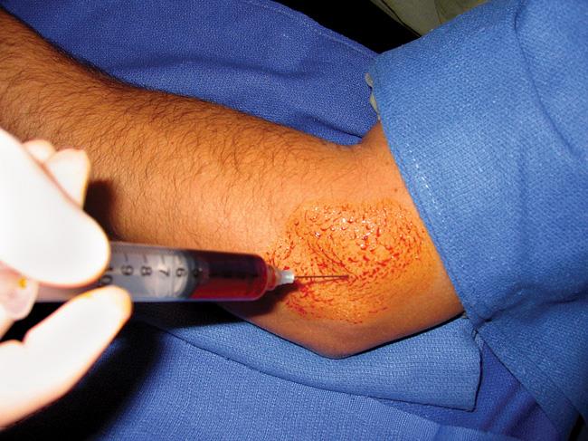
Platelet-rich plasma. Platelet-rich plasma (PRP) is a biological treatment designed to improve the biologic environment of the tissue. This involves obtaining a small sample of blood from the arm and centrifuging it (spinning it) to obtain platelets from the solution. Platelets are known for their high concentration of growth factors, which can be injected into the affected area. While some studies about the effectiveness of PRP have been inconclusive, others have shown promising results.
Extracorporeal shock wave therapy. Shock wave therapy sends sound waves to the elbow. These sound waves create “microtrauma” that promotes the body’s natural healing processes. Shock wave therapy is considered experimental by many doctors, but some sources show it can be effective.
Equipment check.If you participate in a racquet sport, your doctor may encourage you to have your equipment checked for proper fit. Stiffer racquets and looser-strung racquets often can reduce the stress on the forearm, which means that the forearm muscles do not have to work as hard. If you use an oversized racquet, changing to a smaller head may help prevent symptoms from recurring.
If your symptoms do not respond after 6 to 12 months of nonsurgical treatments, your doctor may recommend surgery.
Most surgical procedures for tennis elbow involve removing diseased muscle and reattaching healthy muscle back to bone.
The right surgical approach for you will depend on a range of factors. These include the scope of your injury, your general health, and your personal needs. Talk with your doctor about the options. Discuss the results your doctor has had, and any risks associated with each procedure.
Open surgery. The most common approach to tennis elbow repair is open surgery. This involves making an incision over the elbow.
Open surgery is usually performed as an outpatient surgery. It rarely requires an overnight stay at the hospital.
Arthroscopic surgery. Tennis elbow can also be repaired using miniature instruments and small incisions. Like open surgery, this is a same-day or outpatient procedure.
Surgical risks. As with any surgery, there are risks with tennis elbow surgery. The most common things to consider include:
Rehabilitation. Following surgery, your arm may be immobilized temporarily with a splint. About 1 week later, the sutures and splint are removed.
After the splint is removed, exercises are started to stretch the elbow and restore flexibility. Light, gradual strengthening exercises are started about 2 months after surgery.
Your doctor will tell you when you can return to athletic activity. This is usually 4 to 6 months after surgery. Tennis elbow surgery is considered successful in 80% to 90% of patients. However, it is not uncommon to see a loss of strength.
Three bones make up the ankle joint:
The tibia and fibula have specific parts that make up the ankle:
Two joints are involved in ankle fractures:
Multiple ligaments help make the ankle joint stable.
Because a severe ankle sprain can feel the same as a broken ankle, every ankle injury should be evaluated by a physician.
Common symptoms for a broken ankle include:
After discussing your medical history, symptoms, and how the injury occurred, your doctor will do a careful examination of your ankle, foot, and lower leg.
If your doctor suspects an ankle fracture, he or she will order additional tests to provide more information about your injury.
X-rays. X-rays are the most common and widely available diagnostic imaging technique. X-rays can show if the bone is broken and whether there is displacement (the gap between broken bones). They can also show how many pieces of broken bone there are. X-rays may be taken of the leg, ankle, and foot to make sure nothing else is injured.
Stress test. Depending on the type of ankle fracture, the doctor may put pressure on the ankle and take a special x-ray, called a stress test. This x-ray is done to see if certain ankle fractures require surgery.
Computed tomography (CT) scan. This type of scan can create a cross-section image of the ankle and is sometimes done to further evaluate the ankle injury. It is especially useful when the fracture extends into the ankle joint.
Magnetic resonance imaging (MRI) scan. These tests provide high resolution images of both bones and soft tissues, like ligaments. For some ankle fractures, an MRI scan may be done to evaluate the ankle ligaments.
A lateral malleolus fracture is a fracture of the fibula.
There are different levels at which that the fibula can be fractured. The level of the fracture may direct the treatment.
You may not require surgery if your ankle is stable, meaning the broken bone is not out of place or just barely out of place. A stress x-ray may be done to see if the ankle is stable. The type of treatment required may also be based on where the bone is broken.
Several different methods are used for protecting the fracture while it heals. ranging from a high-top tennis shoe to a short leg cast. Some physicians let patients put weight on their leg right away, while others have them wait for 6 weeks.
You will see your physician regularly to repeat your ankle x-rays to make sure the fragments of your fracture have not moved out of place during the healing process.
If the fracture is out of place or your ankle is unstable, your fracture may be treated with surgery. During this type of procedure, the bone fragments are first repositioned (reduced) into their normal alignment. They are held together with special screws and metal plates attached to the outer surface of the bone. In some cases, a screw or rod inside the bone may be used to keep the bone fragments together while they heal.
A medial malleolus fracture is a break in the tibia, at the inside of the lower leg. Fractures can occur at different levels of the medial malleolus.
Medial malleolar fractures often occur with a fracture of the fibula (lateral malleolus), a fracture of the back of the tibia (posterior malleolus), or with an injury to the ankle ligaments.
If the fracture is not out of place or is a very low fracture with very small pieces, it can be treated without surgery.
A stress x-ray may be done to see if the fracture and ankle are stable.
The fracture may be treated with a short leg cast or a removable brace. Usually, you need to avoid putting weight on your leg for approximately 6 weeks.
You will need to see your physician regularly for repeat x-rays to make sure the fracture does not change in position.
If the fracture is out of place or the ankle is unstable, surgery may be recommended.
In some cases, surgery may be considered even if the fracture is not out of place. This is done to reduce the risk of the fracture not healing (called a nonunion), and to allow you to start moving the ankle earlier.
A medial malleolus fracture can include impaction or indenting of the ankle joint. Impaction occurs when a force is so great it drives the end of one bone into another one. Repairing an impacted fracture may require bone grafting. This graft acts as a scaffolding for new bone to grow on, and may lower any later risk of developing arthritis.
Depending on the fracture, the bone fragments may be fixed using screws, a plate and screws, or different wiring techniques.
A posterior malleolus fracture is a fracture of the back of the tibia at the level of the ankle joint.
In most cases of posterior malleolus fracture, the lateral malleolus (fibula) is also broken. This is because it shares ligament attachments with the posterior malleolus. There can also be a fracture of the medial malleolus.
Depending on how large the broken piece is, the back of the ankle may be unstable. Some studies have shown that if the piece is bigger than 25% of the ankle joint, the ankle becomes unstable and should be treated with surgery.
It is important for a posterior malleolus fracture to be diagnosed and treated properly because of the risk for developing arthritis. The back of the tibia where the bone breaks is covered with cartilage. Cartilage is the smooth surface that lines a joint. If the broken piece of bone is larger than about 25% of your ankle, and is out of place more than a couple of millimeters, the cartilage surface will not heal properly and the surface of the joint will not be smooth. This uneven surface typically leads to increased and uneven pressure on the joint surface, which leads to cartilage damage and the development of arthritis.
If the fracture is not out place and the ankle is stable, it can be treated without surgery.
Treatment may be with a short leg cast or a removable brace. Patients are typically advised not to put any weight on the ankle for 6 weeks.
If the fracture is out of place or if the ankle is unstable, surgery may be offered.
Different surgical options are available for treating posterior malleolar fractures. One option is to have screws placed from the front of the ankle to the back, or vice versa. Another option is to have a plate and screws placed along the back of the shin bone.
“Bi” means two. “Bimalleolar” means that two of the three parts or malleoli of the ankle are broken. (Malleoli is plural for malleolus.)
In most cases of bimalleolar fracture, the lateral malleolus and the medial malleolus are broken and the ankle is not stable.
A “bimalleolar equivalent” fracture means that in addition to one of the malleoli being fractured, the ligaments on the inside (medial) side of the ankle are injured. Usually, this means that the fibula is broken along with injury to the medial ligaments, making the ankle unstable.
A stress test x-ray may be done to see whether the medial ligaments are injured.
Bimalleolar fractures or bimalleolar equivalent fractures are unstable fractures and can be associated with a dislocation.
These injuries are considered unstable and surgery is usually recommended.
Nonsurgical treatment might be considered if you have significant health problems, where the risk of surgery may be too great, or if you usually do not walk.
Immediate treatment typically includes a splint to immobilize the ankle until the swelling goes down. A short leg cast is then applied. Casts may be changed frequently as the swelling subsides in the ankle.
You will need to see your physician regularly to repeat your x-rays to make sure your ankle remains stable.
In most cases, Weightbearing is not be allowed for 6 weeks. After 6 weeks, the ankle may be protected by a removable brace as it continues to heal.
Surgical treatment is often recommended because these fractures make the ankle unstable.
Lateral and medial malleolus fractures are treated with the same surgical techniques as written above for each fracture listed.
“Tri” means three. Trimalleolar fractures means that all three malleoli of the ankle are broken. These are unstable injuries and they can be associated with a dislocation.
These injuries are considered unstable and surgery is usually recommended.
As with bimalleolar ankle fractures, nonsurgical treatment might be considered if you have significant health problems, where the risk of surgery may be too great, or if you usually do not walk.
Nonsurgical treatment is similar to bimalleolar fractures, as described above.
Each fracture can be treated with the same surgical techniques as written above for each individual fracture.
The syndesmosis joint is located between the tibia and fibula, and is held together by ligaments. A syndesmotic injury may be just to the ligament — this is also known as high ankle sprain. Depending on how unstable the ankle is, these injuries can be treated without surgery. However, these sprains take longer to heal than the normal ankle sprain.
In many cases, a syndesmotic injury includes both a ligament sprain and one or more fractures. These are unstable injuries and they do very poorly without surgical treatment.
Your physician may do a stress test x-ray to see whether the syndesmosis is injured.
Because there is such a wide range of injuries, there is also a wide range of how people heal after their injury. It takes at least 6 weeks for the broken bones to heal. It may take longer for the involved ligaments and tendons to heal.
As mentioned above, your doctor will most likely monitor the bone healing with repeated x-rays. This is typically done more often during the first 6 weeks if surgery is not chosen.
Pain after an injury or surgery is a natural part of the healing process. Your doctor and nurses will work to reduce your pain, which can help you recover faster.
Medications are often prescribed for short-term pain relief after surgery or an injury. Many types of medicines are available to help manage pain, including opioids, non-steroidal anti-inflammatory drugs (NSAIDs), and local anesthetics. Your doctor may use a combination of these medications to improve pain relief, as well as minimize the need for opioids.
Be aware that although opioids help relieve pain after surgery or an injury, they are a narcotic and can be addictive. It is important to use opioids only as directed by your doctor. As soon as your pain begins to improve, stop taking opioids. Talk to your doctor if your pain has not begun to improve within a few days of your treatment.
Rehabilitation is very important regardless of how an ankle fracture is treated.
When your physician allows you to start moving your ankle, physical therapy and home exercise programs are very important. Doing your exercises regularly is key.
Eventually, you will also start doing strengthening exercises. It may take several months for the muscles around your ankle to get strong enough for you to walk without a limp and to return to your regular activities.
Again, exercises only make a difference if you actually do them.
Your specific fracture determines when you can start putting weight on your ankle. Your physician will allow you to start putting weight on your ankle when he or she feels your injury is stable enough to do so.
It is very important to not put weight on your ankle until your physician says you can. If you put weight on the injured ankle too early, the fracture fragments may move or your surgery may fail and you may have to start over.
It is very common to have several different kinds of things to wear on the injured ankle, depending on the injury.
Initially, most ankle fractures are placed in a splint to protect your ankle and allow for the swelling to go down. After that, you may be put into a cast or removable brace.
Even after the fracture has healed, your physician may recommend wearing an ankle brace for several months while you are doing sporting activities.
People who smoke, have diabetes, or are elderly are at a higher risk for complications after surgery, including problems with wound healing. This is because it may take longer for their bones to heal.
Without surgery, there is a risk that the fracture will move out of place before it heals. This is why it is important to follow up with your physician as scheduled.
If the fracture fragments do move out of place and the bones heal in that position, it is called a “malunion.” Treatment for this is determined by how far out of place the bones are and how the stability of the ankle joint is affected.
If a malunion does occur or if your ankle becomes unstable after it heals, this can eventually lead to arthritis in your ankle.
General surgical risks include:
Risks from the surgical treatment of ankle fractures include
Although most people return to normal daily activities, except for sports, within 3 to 4 months, studies have shown that people can still be recovering up to 2 years after their ankle fractures. It may take several months for you to stop limping while you walk, and before you can return to sports at your previous competitive level. Most people return to driving within 9 to 12 weeks from the time they were injured.
The Achilles tendon is a strong fibrous cord present behind the ankle that connects the calf muscles to the heel bone. It is used when you walk, run and jump. The Achilles tendon ruptures most often in athletes participating in sports that involve running, pivoting and jumping. Recreational sports that may cause Achilles rupture include tennis, football, basketball and gymnastics.
When the Achilles tendon ruptures, you will experience severe pain in the back of your leg above your heel, swelling, stiffness, and difficulty standing on tiptoe and moving the leg when walking. A popping or snapping sound may be heard when the injury occurs.
Your doctor diagnoses the rupture based on symptoms, history of the injury and physical examination. Your doctor may also feel a gap or depression in the tendon, just above the heel bone. Your doctor will gently squeeze the calf muscles. If the Achilles tendon is intact, there will be flexion movement of the foot, if it is ruptured, there will be no movement observed.
Achilles tendon rupture is treated using nonsurgical methods or surgery. Nonsurgical treatment involves wearing a cast or special brace which lifts your heel, allowing the tendon to heal. Surgical procedure involves opening the skin and suturing the torn tendon together. Surgery helps to decrease the recurrence of the Achilles tendon in comparison to the nonsurgical treatment. With either treatment, physical therapy is recommended to improve the strength and flexibility of the leg muscles and the Achilles tendon.
To help prevent an Achilles tendon injury, it is a good practice to perform stretching and warm-up exercises before participating in any exercises or sports activities. Gradually increase the intensity and length of time of the activity. Muscle conditioning may help to strengthen the muscles in the body.
Ankle instability surgery is performed to treat an unstable ankle and involves the repair or replacement of a torn or stretched ligament.
There are two types of ankle instability surgery:
Ankle instability is a chronic condition characterized by a recurrent slipping of the outer side of the ankle. Instability is generally noticed during movement of the ankle joint but can also occur during standing as well.
Symptoms include the following:
Ankle Instability usually results from repeated ankle sprains. Inadequate healing of a sprained ligament or incomplete rehabilitation of the affected ligament can result in instability. Recurrent injury to the ligaments further weakens them and aggravates the instability which predisposes to the development of additional ankle problems.
Surgery is recommended in patients with a high degree of ankle instability and in those who have failed to respond to non-surgical treatments.
Ankle instability surgery involves the repair or reconstruction of the injured ankle ligaments. Ankle-instability surgeries can be categorized into either anatomic repair or non-anatomic repair, also called reconstructive tenodesis.
Anatomic repair involves reconstruction of the stretched or torn ligaments. The surgery is performed under epidural anesthesia. Your surgeon makes an incision on the ankle to expose the damaged joint and ligaments. The joint capsule and ligaments are examined and the edges of the torn ligament are shortened and repaired with sutures. The ends may be overlapped and then sutured to strengthen the ligament. Your surgeon then covers the repaired ligament with the extensor retinaculum, a dense band of connective tissue, to reinforce the ligament further. Range of motion is evaluated; the incision is closed and a sterile bandage is applied.
Reconstructive tenodesis is a tendon transfer procedure that uses your own tendon or a cadaver tendon as a graft to replace the damaged tendon. The surgery is performed under epidural anesthesia. Your surgeon makes an incision on your ankle. Drill holes are created where the damaged ligament normally attaches to the lower end of the fibula (calf bone) on one side and the talus (anklebone) on the other end. Your surgeon then harvests the peroneus brevis muscle tendon, found on the outer edge of the small toe, and weaves it through the drill holes to form a ligament complex. Range of motion is evaluated; the incision is closed and a sterile bandage is applied.
After surgery, your foot will be immobilized with a cast or splint. You will be provided crutches to avoid bearing weight on the operated ankle. Your doctor will remove the splint and provide a removable boot to be worn for 2 to 4 weeks. Physical therapy will be initiated to strengthen your joint and improve range of motion. Complete recovery may take 10 to 12 weeks.
The advantages of the anatomic repair include:
The disadvantage of the anatomic repair includes:
The advantages of the nonanatomic repair include:
The disadvantages of the nonanatomic repair procedures include:
Does not preserve the peroneus brevis, an important structure for the ankle’s dynamic stability
As with all surgical procedures, ankle instability surgery may be associated with certain complications including:
Your shoulder joint is a ball and socket joint made up of the upper arm bone, the shoulder blade and the collarbone. The head of the upper arm bone fits into the socket of the shoulder joint known as the glenoid cavity. The outer edge of the glenoid is surrounded by a strong fibrous tissue called the labrum.
A superior labrum anterior and posterior tear or SLAP tear is an injury to the labrum. This injury may also involve the biceps tendon, which is attached to the top part of the labrum. The injury occurs from repeated use of the shoulder while throwing or a fall onto the shoulder. A SLAP tear can be treated through an arthroscopic surgical procedure called a SLAP repair.
A SLAP repair is indicated to treat the torn labrum of the shoulder socket when conservative treatments measures such as NSAID’s (non-steroidal anti-inflammatory medications) and Physical Therapy do not relieve the symptoms of a SLAP tear.
A SLAP repair is a minimally invasive surgery using an arthroscope, a tube with a light and camera on the end that projects images onto a monitor for your surgeon to view inside your joint. The type of SLAP repair depends on the type of tear involved and will be determined once your surgeon views the joint. The procedure is performed under general anesthesia and nerve block.
After the procedure, your arm will be placed in a sling for the first 3 weeks to immobilize the shoulder joint and you will be advised to restrict active motion of your shoulder for about 6 weeks. You will be instructed take your pain medications and to apply ice packs to control swelling and pain. The plaster strips over the wounds should be kept dry until the wounds heal. Through physical therapy, you can slowly regain motion and strengthen the shoulder. You can resume sport activities after consulting with your physical therapist and surgeon. You should avoid driving for a few weeks after the surgery.
A SLAP repair involves the reattaching of the labrum to its normal anatomical position; thereby restoring the anatomy of the shoulder. The procedure also allows normal functioning of the previously damaged labrum and biceps attachment.
As with any surgical procedure, SLAP repair involves certain potential risks and complications including
The shoulder joint is a ball and socket joint. A ‘ball’ at the top of the upper arm bone (the humerus) fits neatly into a ‘socket’, called the glenoid, which is part of the shoulder blade (scapula). The labrum is a ring of fibrous cartilage surrounding the glenoid, which helps in stabilizing the shoulder joint. The biceps tendon is attached inside the shoulder joint at the superior labrum of the joint. The biceps tendon is a long cord-like structure which attaches the biceps muscle to the shoulder and helps to stabilize the joint.
Traumatic injury to the shoulder or overuse of the shoulder by excessive throwing or weightlifting can cause a labral tear. In addition, the ageing process may weaken the labrum, leading to injury secondary to wear and tear.
A shoulder labral tear injury can cause symptoms such as pain, a catching or locking sensation, decreased range of motion and joint instability.
The most common types of labral tears include:
Your doctor may suspect a labral tear based on your symptoms and medical history. The doctor will inquire about your pain and history of injury. Several physical tests will be performed by your doctor to evaluate the range of motion and stability of your shoulder. X-rays may be used to rule out other conditions. Your doctor may also order a computed tomography (CT) scan or magnetic resonance imaging (MRI) scan, with a contrast medium, to determine the presence of tears. Diagnosis of a labral tear can also be confirmed through shoulder arthroscopy.
Your doctor may start with conservative approaches such as prescribing anti-inflammatory medications and advise rest to relieve symptoms. Rehabilitation exercises may be recommended to strengthen the rotator cuff muscles. If the symptoms do not resolve with these conservative measures, your doctor may recommend arthroscopic surgery.
During arthroscopic surgery for SLAP tears, your surgeon examines the labrum and the biceps tendon. If the damage is confined to the labrum without involving the tendon, then the torn flap of the labrum will be removed. In cases where the tendon is also involved or if there is detachment of the tendon, absorbable wires or sutures will be used to repair and reattach the tendon.
Bankart lesion is repaired by a Bankart operation. In this procedure, the Bankart tear is repaired by reattaching the labrum and the capsule to the anterior margin of the glenoid cavity. Your surgeon makes a few small incisions around the joint. Through one incision, an arthroscope is inserted into the shoulder to visualize the inside of the shoulder joint. Other surgical instruments are inserted through the other incision to re-attach the labrum to the glenoid with the help of sutures or anchoring devices. The arthroscope and surgical instruments are removed and the incisions are closed.
Following the surgery, your shoulder is immobilized with a sling for a few days. To control pain and swelling, your physical therapist may use ice, electrical stimulation, massage therapy, and other hands-on treatments. Passive range of motion exercises are also initiated in the Post-operative phase. Active range of motion exercises are started about 6 weeks after the repair, to regain your shoulder movement. Athletes can return to sports in about three months.
The risks associated with a labral repair include:
The anterior cruciate ligament is one of the major stabilizing ligaments in the knee. It is a strong rope like structure located in the center of the knee running from the femur to the tibia. When this ligament tears unfortunately, it does not heal and often leads to the feeling of instability in the knee.
An ACL injury most commonly occurs during sports that involve twisting or overextending your knee. An ACL can be injured in several ways:
When you injure your ACL, you might hear a loud “pop” sound and you may feel the knee buckle. Within a few hours after an ACL injury, your knee may swell due to bleeding from vessels within the torn ligament. You may notice that the knee feels unstable or seems to give way, especially when trying to change direction on the knee.
An ACL injury can be diagnosed with a thorough physical examination of the knee and diagnostic tests such as X-rays, MRI scans and arthroscopy. X-rays may be needed to rule out any fractures. In addition, your doctor will often perform the Lachman’s test to see if the ACL is intact. During a Lachman test, knees with a torn ACL may show increased forward movement of the tibia and a soft or mushy endpoint compared to a healthy knee.
Pivot shift test is another test to assess ACL tear. During this test, if the ACL is torn, the tibia will move forward when the knee is completely straight and as the knee bends past 30° the tibia shifts back into correct place in relation to the femur.
The goal of ACL reconstruction surgery is to tighten your knee and to restore its stability.
Anterior cruciate ligament reconstruction hamstring method is a surgical procedure to replace the torn ACL with part of the hamstring tendon taken from the patient’s leg. The Hamstring is the muscle located on the back of your thigh. The procedure is performed under general anesthesia.
Your surgeon will make two small cuts about 1/4-inch-long around your knee. An arthroscope, a tube with a small video camera on the end is inserted through one incision to see the inside of the knee joint. Along with the arthroscope, a sterile solution is pumped into the joint to expand it enabling the surgeon to have a clear view and space to work inside the joint.
The knee is bent at right angles and the hamstring tendons felt. A small incision is made over the hamstring tendon attachment to the tibia and the two tendons are stripped off the muscle and the graft is prepared. The torn ACL will be removed and the pathway for the new ACL is prepared. The arthroscope is reinserted into the knee joint through one of the small incisions. Small holes are drilled into the upper and lower leg bones where these bones come together at the knee joint. The holes’ form tunnels in your bone to accept the new graft. Then the graft is pulled through the predrilled holes in the tibia and femur. The new tendon is then fixed into the bone with screws to hold it into place while the ligament heals into the bone.
The incisions are then closed with sutures and a dressing is placed.
Possible risks and complications associated with ACL reconstruction with hamstring method include:
Following the surgery, rehabilitation begins immediately. A physical therapist will teach you specific exercises to be performed to strengthen your leg and restore knee movement. Avoid competitive sports for 5 to 6 months to allow the new graft to incorporate into the knee joint.
Anterior cruciate ligament reconstruction is a very common and successful procedure. It is usually indicated in patients wishing to return to an active lifestyle especially those wishing to play sports involving running and twisting. Anterior cruciate ligament injury is a common knee ligament injury. If you have injured your ACL, surgery may be needed to regain full function of your knee.
Meniscus tear is the commonest knee injury in athletes, especially those involved in contact sports. A sudden bend or twist in your knee cause the meniscus to tear. This is a traumatic meniscus tear. Elderly people are more prone to degenerative meniscal tears as the cartilage wears out and weakens with age. The two wedge-shaped cartilage pieces’ present between the thighbone and the shinbone are called meniscus. They stabilize the knee joint and act as “shock absorbers”.
Torn meniscus causes pain, swelling, stiffness, catching or locking sensation in your knee making you unable to move your knee through its complete range of motion.
Your orthopedic surgeon will examine your knee, evaluate your symptoms, and medical history before suggesting a treatment plan. The treatment depends on the type, size and location of tear as well your age and activity level. If the tear is small with damage in only the outer edge of the meniscus, nonsurgical treatment may be sufficient. However, if the symptoms do not resolve with nonsurgical treatment, surgical treatment may be recommended.
Knee arthroscopy is the commonly recommended surgical procedure for meniscal tears. The surgical treatment options include meniscus removal (meniscectomy), meniscus repair, and meniscus replacement. Surgery can be performed using arthroscopy where a tiny camera will be inserted through a tiny incision which enables the surgeon to view inside of your knee on a large screen and through other tiny incisions, surgery will be performed. During meniscectomy, small instruments called shavers or scissors may be used to remove the torn meniscus. In arthroscopic meniscus repair the torn meniscus will be pinned or sutured depending on the extent of tear.
Meniscus replacement or transplantation involves replacement of a torn cartilage with the cartilage obtained from a donor or a cultured patch obtained from laboratory. It is considered as a treatment option to relieve knee pain in patients who have undergone meniscectomy.